Restoration of an Abnormal Cervical Lordosis Using the DENNEROLL: A CBP® Case Report
 Monday, April 12, 2010 at 11:30PM
 | Brian Paris, BS, DC | Deed E. Harrison, DC |
|
Advanced CBP Fellow Private Practice Rockville, MD |
President CBP Seminars, Inc. Vice President CBP Non-Profit, Inc. Chair PCCRP Guidelines Editor—AJCC |
Introduction
In surgical1,2 and non-surgical literature3-5, it has been found that patients with less cervical lordosis have statistically significant increases in neck, upper thoracic, and shoulder pain and likely overall poorer health outcomes. The following case report suggests that the loss of cervical lordosis, forward head carriage, spinal arthritis and disc disease (S.A.D.D.), with concomitant sympomatology is consistent with current literature relating to cervical lordosis and pain.1-5
Case History Key Features
A 59 year old female present to the practice of one of the authors (BP) seeking help for recurrent and chronic dizziness, neck and shoulder pain and stiffness, left arm tingling, sleep deprivation, and generalized fatigue. Her initial visit was on 10-7-2009 and she indicated that the current episode of her ailments had been present for at least the previous 2 months.
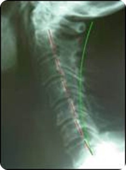
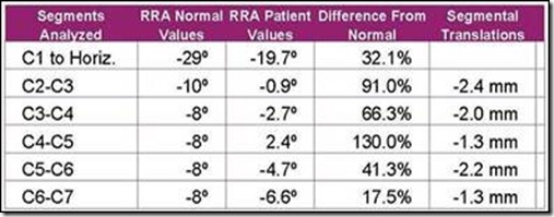
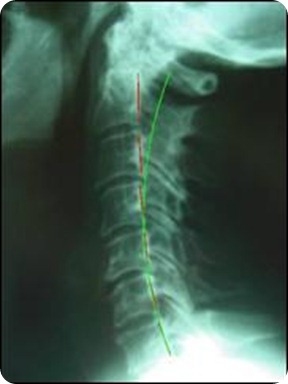
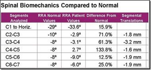
Cervical radiographs consisting of a neutral lateral and anterior-posterior were obtained. The lateral cervical radiograph revealed signs of spinal arthritis and disc disease (S.A.D.D) increasing in severity at the C5-C7 levels with possible rheumatoid changes in the upper cervical region. The patient had a large forward head carriage (58 mm from C2-C7) and a 70% reduction of the normal cervical lordosis. Radiographic analytical measurements and comparison to normal values were performed with the PostureRay® computerized software system from PostureCo. See Figure 1.
Figure 1. Patient Pre-Lateral Cervical Radiograph and segmental alignments relative to ideal values. The green semi-circular curvature is the ideal curvature proposed by Harrison et al and the dashed red line represents the path of the patient’s posterior vertebral bodies and visually depicts the amount of displacement. The initial cervical lordosis demonstrated a 70% reduction, 12.6° from C2-C7 using the Harrison Posterior Tangent method.6,7 The initial forward translation was 58 mm; using posterior superior body corner of C2 relative to a vertical line originating at the posterior inferior body corner of C7.
CBP Mirror Image® Interventions
The patient was recommended and agreed to a treatment plan of spinal correction using CBP Technique mirror image adjustment, exercise, and traction methods. The treatment frequency was 3 times per week for 40 visits over approximately 13 weeks. The Patient presented to and actively participated at all appointments. Each visit consisted of mirror image adjusting, mirror image exercises and the Denneroll cervical orthotic to improve the cervical lordosis and reduce abnormal posture displacements.
- Mirror Image Adjustments
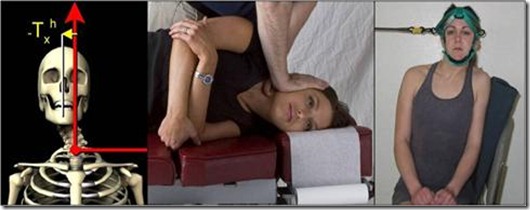
Beginning on 10-9-09, the patient was administered mirror image adjustments to correct Right Head Translation and Anterior Head Translated postures. See Figures 2 and 3.
- Mirror Image Exercises
Active rehabilitative care was administered to the patient beginning on 10-9-09. Since the patient’s abnormal posture was found to be Right Head Translation (-TxH) and Anterior Head translation (+TzH), the patient began mirror image exercises in left head translation to correct right head translation and posterior head translation without extension due to dizziness. See Figures 2 and 3.
Figure 2. The patient presented with the abnormal posture of Right Head Translation (-TxH). Mirror image adjustments were given in Left Head Translation. Mirror image exercises in Left Head Translation were given to the patient as part of CBP Active Rehabilitative care.
Figure 3. The patient presented with the abnormal posture of Anterior Head Translation (+TzH). Mirror image adjustments were given in Posterior Head Translation with extension to improve the lordosis and reduce forward head translation. Mirror image exercises in Posterior Head Translation (extension exercises caused increased dizziness) were given to the patient as part of CBP Active Rehabilitative care.
- The Denneroll Cervical Orthotic Intervention
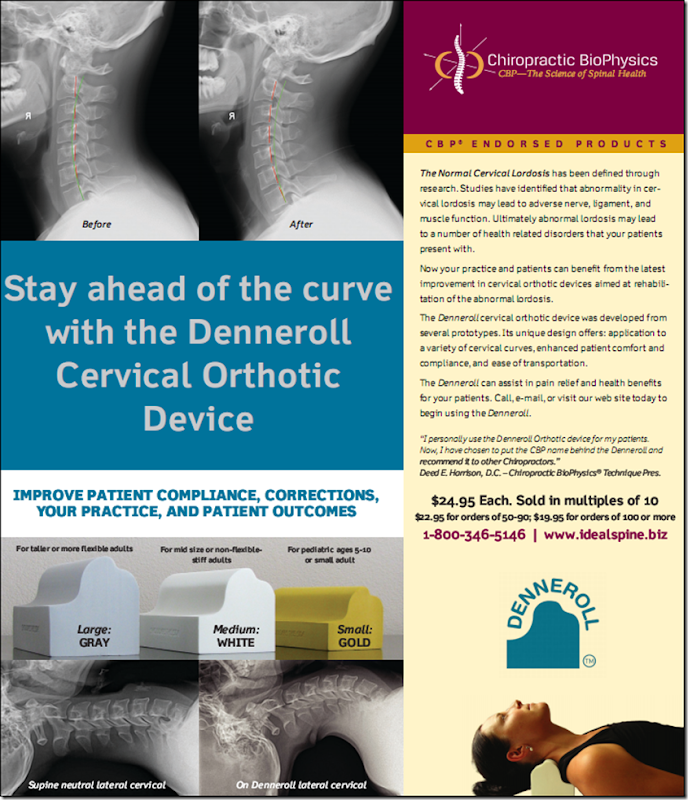
The patient experienced considerable difficulty in performing passive and active cervical extension. Accordingly, the more advanced types of CBP in office traction methods could not be performed by the patient.1 Thus, the Denneroll cervical orthotic device (Figure 4) was provided to the patient as the sole method of in office cervical corrective traction-stretching. Denneroll (adult small size) corrective stretching began on 10/19/2009 and continued for a total of 34 treatment sessions. Patient time started at 3 minutes per session and then increased up to 10 minutes per Denneroll session each visit in the office.
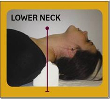
For the current patient, the Denneroll was placed in the upper thoracic/lower cervical region (Figure 4). This placement of the Denneroll will cause significant posterior head translation, will increase the upper thoracic curve if the large device is used (this is why the small device was used herein), and will increase the overall cervical lordosis.
Figure 4. Denneroll corrective orthotic application in the lower cervical region. This lower neck placement is for abnormal cervical curvatures having:
· Normal or a mild loss of the upper thoracic kyphosis;
· Loss of the mid-lower cervical curve;
Anterior head translation of approximately ≤ 40mm
Figure 5. Patient Post-Lateral Cervical Radiograph and segmental alignments relative to ideal values. The green semi-circular curvature is the ideal curvature proposed by Harrison et al and the dashed red line represents the path of the patient’s posterior vertebral bodies and visually depicts the amount of displacement. The Follow-up cervical lordosis demonstrated a 56% reduction, 18° from C2-C7 using the Harrison Posterior Tangent method.6,7 The initial forward translation was 19 mm; using posterior superior body corner of C2 relative to a vertical line originating at the posterior inferior body corner of C7.
Re-Examinations:
In addition to the initial examination, 2 follow-up evaluations were performed over the course of the 40 sessions. At each examination, structural and functional responses to care were evaluated and patient symptoms were recorded and monitored using the Neck Disability Index and the Rand 36- Health Status Questionnaire. See Table 1. The brief examination findings are summarized here:
- 11/12/2009: Decreased dizziness; thinking more clearly; energy level (fatigue) same; left arm tingling improvingànow only intermittent;
- 12/14/2009: Infrequent bouts of dizziness, only occasional tingling in left arm;
- 1/13/2010: No reports of dizziness, occasional tingling in left arm.
- Overall Improvements: Significant health improvements were noted by patient since beginning treatment: “No dizziness”; “more clear-headed”. At most recent follow-up, dizziness had not returned. Only occasional tingling in left hand was reported and the patient has elected to continue with a 2nd phase of CBP Corrective Care.
Table 1. Initial and Follow-up Neck Disability and Rand-36 Questionnaire results.
|
Questionnaire |
Initial Exam 11-12-2009 |
Re-Exam 1-13-2010 |
|
Neck Disability |
22% Pain interference with ADL’s |
12% Pain interference with ADL’s |
|
Rand-36-Health Status Questionnaire 12-14-09 1-13-10 |
||
|
Physical Function |
60 |
75 |
|
Social Function |
62.5 |
75 |
|
Role Physical |
100 |
100 |
|
Role Emotional |
0 |
100 |
|
Mental Health |
64 |
72 |
|
Energy-Fatigue |
45 |
55 |
|
Pain |
45 |
67.5 |
|
Health Perception |
77 |
52 |
Discussion
The Denneroll orthotic applies a passive 3-point bending force to the cervical spine that is generally well tolerated and is most consistent with the Pope-2-way type of in office corrective traction force.6The Denneroll is available in 2 adult sizes (adult large and adult small) and the adult small device was used for the present patient. The Denneroll size and placement of the device must be consistent with both the shape of the cervical curve and the amount/type of sagittal head translation correction that is desired for the given patient.
In the current case report, the combination of CBP mirror image methods resulted in improvement of cervical spine vertebral subluxation towards normal alignment. The Denneroll Orthotic for cervical lordosis corrective traction-stretching was the only type of CBP Traction utilized herein. Thus, it appears that the Denneroll Orthotic device assisted in cervical spine correction and improvement in chronic patient symptoms, disability, and altered health when applied in combination with mirror image adjustments and active mirror image exercises.
We will continue to test the Denneroll device in appropriate patient cases and provide the results in future articles.
References
- Lowery G. Three-dimensional screw divergence and sagittal balance: a personal philosophy relative to cervical biomechanics. Spine: State of the Art Reviews 1996;10:343-356.
- Kawakami M, Tamaki T, Yoshida M, Hayashi N, Ando M, Yamada H. Axial symptoms and cervical alignments after cervical anterior spinal fusion for patients with cervical myelopathy. J Spinal Disorders 1999;12:50-56.
- Kai Y, Oyama M, Kurose S, et al. Traumatic thoracic outlet syndrome. Orthop Traumatol 1998;47:1169-1171.
- Harrison DD, Harrison DE, Janik TJ, Cailliet R, Haas JW, Ferrantelli J, Holland B. Modeling of the Sagittal Cervical Spine as a Method to Discriminate Hypo-Lordosis: Results of Elliptical and Circular Modeling in 72 Asymptomatic Subjects, 52 Acute Neck Pain Subjects, and 70 Chronic Neck Pain Subjects. Spine 2004; 29:2485-2492.
- McAviney J, Schulz D, Richard Bock R, Harrison DE, Holland B. Determining a clinical normal value for cervical lordosis. J Manipulative Physiol Ther 2005;28:187-193.
- Harrison DE, Harrison DD, Haas JW. CBP Structural Rehabilitation of the Cervical Spine. Chapters 2 & 6. CBP Seminars, 2002; pgs:147-151. ISBN 0-9721314-0-X.
- Harrison DD, et al. Spine 2004; 29:2485-2492.
 CBP Seminars | Comments Off |
