Sunday
Jan112015

Treating the Elderly with Chiropractic BioPhysics® or CBP® Technique Methods

Jason W. Haas, DC

Private Practice Windsor, CO

Many practitioners are surprised to find out the extent of Chiropractic BioPhysics or CBP Technique methods we employ to treat elderly patients in our facility.  Some colleagues feel that the elderly patients may not be a candidate for treatment due to the fact that they are older, frailer, can have extensive degeneration or many comorbidities.1-3 These doctors are afraid to attempt to change their spine and posture because the problem is long-standing or they fear they may injure the patient.  However, in our clinical experience treating over 6500 patients in the last eleven years, we have found that the elderly patients respond very well to gentle application of CBP methods and astute clinicians will find that they can have a tremendous positive effect on a patient’s posture, pain, quality of life and overall health.

                  Most of our elderly patients come to us for painful conditions that they have not been able to find relief from using traditional treatments of drugs, surgery or even traditional chiropractic and physical therapy.  These patients are often discouraged with healthcare in general because they are finding that the time they spend with their doctor is less and less and the outcomes of the medicines they are prescribed are less and less effective. This can create a situation of frustration that a well-trained receptionist must contend with in order to get the elderly person feeling comfortable in the office.  A great tour of the office and a compassionate staff can show these folks that the CBP experience will likely be completely different from other chiropractors, medical doctors and other healthcare providers

Our Intake Process & General Exam Procedures

                  Once they see that we offer a different approach to spinal care, we can move them through the process of intake.  The examination is similar to younger patients but may require more time due to a longer health history and multiple concomitant health concerns/conditions.  We will always perform a thorough health history, all pertinent orthopedic and neurological testing, blood analysis if necessary, CBP structural evaluation including spine and extremity films when indicated, Posture Screen™ digital postural analysis and all pertinent outcome measures (all patients receive the SF-36, pain and disability questionnaires), and digital range of motion and strength testing.

                  Once we have the data for our initial assessment we make a determination as to whether they are candidates for CBP Structural Rehabilitation; yes there are some cases that we won't perform CBP care with due to contra-indications.  This assessment involves looking at the evaluated spine films with PostureRay™, and determining if there are any areas that would not respond well to conservative care, and CBP care especially.  If there are areas of suspected instability we will take flexion/extension films or send to my friend Evan Katz,DC for DMX.  Once possible contraindications have been ruled out, we explain the importance of improving sagittal and coronal balance with the patient and discuss the limitations that the care may have.  The discussions on limitations are crucially important because the elderly patients must be aware that osteophytes, disc and ligament degeneration and severe structural abnormalities will likely not return to normal as a result of the care we are providing.  They must know that the goals are better range of motion and strength, better posture (coronal and sagittal) and improvements measured on their outcome measures (SF-36, NDI, etc.) If a patient has an unrealistic expectation of being “fixed” and getting back to completely normal, it’s important to inject some reality and sometimes limit expectations.

Sagittal Plane Alignment & Health Concerns in the Elderly

                  The sagittal plane alignment of the spine and posture and it's connection to human health and longevity is becoming one of the most widely researched topics in spine care today.4-10 The sagittal plane alignment of the spine and posture in the elderly has been found to correlate to the following health disorders:

  1. Increased risk of spinal compression fractures;4
  2. Increased low back pain and more severe pain;5
  3. Decreased mobility and increased risk of falls leading to fractures;6,7,8
  4. Increased risk of going into a care giving facility and not being able to take care of one's self in normal activities of daily living;9
  5. Increased knee or patellar femoral pain;10
  6. Increased disability and impairment due to pain;11
  7. Possible increase risk of early death compared to age match corhorts.12,13

For example, of a few of these important studies are reviewed here. Kobayashi and colleagues4 prospectively followed 100 subjects aged 61.9 yrs of age for an additional 12 years (indicating they were about 74 years at follow up). Full spine radiographs were ascertained at initial and long-term follow up in an attempt to identify if sagittal plane radiographic alignment variables play a role in the risk for developing new vertebral compression fractures. In both univariate and multivariate analysis, reductions in lumbar lordosis (Cobb L1-L5) and thoracic kyphosis (Cobb T4-T12) increased the relative risk of developing a new vertebral body compression fracture. Significantly, even curves one standard deviation below the mean value showed statistically significant increased relative risks (RR 3.06). Their4 most statistically significant model was multi-variate including pre-existing compression fractures with both the lumbar lordosis and thoracic kyphosis decreased (RR 8.64). Kobayashi and colleagues4 suggested that flattened curves reduce the shock absorbing capability of the sagittal curves, increasing the dynamic loads on the vertebral bodies thus increasing the risk of fractures.

                  In a prospective study of 253 chronic LBP patients matched by age and physical characteristics to 253 normal controls between the ages of 50-85 years, Tsuji et al5 found a reduced L1-S1 lordosis in the chronic LBP group. Of primary importance, lumbar lordosis was inversely correlated with pain intensity on a visual analog scale (p= 0.025). In other words, as the lumbar lordosis decreased, the pain intensity of the subject was increased.5

                  Recently, Kamitami et al9, studied the spinal posture in 804 participants (65–94 yrs of age) who were initially independent in their activities of daily living (ADLL) at baseline. These participants were followed for a 4.5-year follow-up period and it was found that 126 (15.7%) of the participants became dependent in their ADLs. Dependence in ADL was defined as admission to a nursing home or need of home assistance to perform basic self care functions. Importantly, inclination of the upper body relative to the pelvis (angle subtended between the vertical and a line joining C7 to the sacrum) was correlated with outcome and lumbar curvature also showed a marginal association. After adjusting for age and sex, it was found that for each 1 unit increase in the quartile of forward inclination that the odds of becoming dependent on ADL's was 1.79 x greater. Indicating that the highest quartile had a risk of 1.79 x 3 = 5.37 times more likely to be dependent. This study is very important for the elderly person wanting to remain able to perform basic care functions.

Chiropractic BioPhysics Mirror Image® Treatment Approach

                  Care of the elderly patient is slightly different in the CBP setting as it requires a gentler; yet still patient centered approach. We are cautious not to use high-velocity manipulation with elderly patients and work primarily on improving their posture with gentle CBP Mirror Image adjustments. The CBP Mirror image exercises are incredibly important in increasing strength and stability of the core postural muscles. We recommend and use the PowerPlate® as this is an exceptional tool for the elderly to perform their Mirror Image exercises while getting maximal benefit from the whole body vibration or acceleration training.

                  The CBP Mirror Imag traction protocols must also be altered for the elderly patients, they must understand why we are using the traction and stressing the analogy that spinal change is a marathon and not a sprint is important to minimize injury.  Most elderly patient in our office have radiculopathy, peripheral neuropathies, disc herniations and osteophytes so the use of distractive traction is often superior to compression traction which can irritate and worsen radiating symptoms.

                  Frequent re-evaluation and re-assessment of the elderly patient is possibly more important than any other age group.  This is due to the fact that they often have significant health complications that ned to be assessed periodically.  In our integrated setting it is easy for them to see our nurse practitioner or medical doctor if new symptoms arise, and for those of you who are not integrated, I would suggest having a close relationship with local providers to manage any health complications that may arise throughout care.

                  Diligent, patient, and compassionate care for the Elderly population can provide this segment with significant health gains, better posture and balance, less pain and a better overall quality of life. IF you would like more information on CBP care and the elderly, please attend any upcoming seminar.

Sample Patient Case:

Age: 87

Initial Complaints:  Low back pain and left leg sciatic pain NRS 8/10

Oswestry:  58% disability. Significant depression due to worsening of pain, poor balance and coordination.

Patient was told by two other chiropractors they could not help her.  She was told by a surgeon that she is not a candidate for surgery due to advanced age.

CBP Treatment:  Mirror Image adjustments prone with the headpiece elevated and instrument adjustment to the spine. Mirror Image exercises; standing with a block in the lower thorax and head and ribcage retraction performed in-office and at home.  Supine thoraco lumbar traction. Fulcrum of traction at thoraco-lumbar junction, 0° angle of pull and leg strapped below the femur heads, sustained for 12 minutes. Cervical Traction consisting of  Pope-2-way type traction with a distractive force of 10 lbs and a finishing front weight of 18 lbs. sustained for 10 minutes.

Total treatments:  30 sessions

Figure 1. Before treatment and after treatment full spine radiographic changes due to CBP mirror image care over the course of 30 visits. Note that the follow up x-rays are taken a minimum of 3 days after the patients last treatment session. Thus, this is not an x-ray immediately after care. In this manner an accurate response to care can be found.

Final Complaints:  Very rare low back pain (NRS2/10)  Sciatica resolved, Depression significantly reduced and the patient states she feels better now than she has in decades. Oswestry: 8%.

Perspective on Patient Outcome:

                  It is my opinion that the improvement in the patient's condition, outcome measures, and self reported ability to function was due to the considerable improvement in the sagittal plane alignment of the patient's thoraco-lumbar curvatures and sagittal-forward balance. The references4-13 provided below provide evidence based support for this anecdotal but clinically obvious statement.


Reference Links

  1. http://www.ncbi.nlm.nih.gov/pubmed/25436061
  2. http://www.ncbi.nlm.nih.gov/pubmed/23307577
  3. http://www.ncbi.nlm.nih.gov/pubmed/25533322
  4. http://www.ncbi.nlm.nih.gov/pubmed/?term=Kobayashi+T+Osteoporos+Int++2008
  5. http://www.ncbi.nlm.nih.gov/pubmed/11479757
  6. http://www.ncbi.nlm.nih.gov/pubmed/21198460
  7. http://www.ncbi.nlm.nih.gov/pubmed/24715607
  8. http://www.ncbi.nlm.nih.gov/pubmed/20480146
  9. http://www.ncbi.nlm.nih.gov/pubmed/?term=Kamitami+2013
  10. http://www.ncbi.nlm.nih.gov/pubmed/12355123
  11. http://www.ncbi.nlm.nih.gov/pubmed/15972617
  12. http://www.ncbi.nlm.nih.gov/pubmed/19451575
  13. http://www.ncbi.nlm.nih.gov/pubmed/15450042
Monday
Dec222014

The Cervical Lordosis in Health and Disease: Literature Review & The Denneroll 'Home Based' Orthotic

Deed E. Harrison, DC President CBP Seminars, Inc. Vice President CBP Non-Profit, Inc. Chair PCCRP Guidelines Editor—AJCC Private Practice-- Eagle, ID, USA

INTRODUCTION

Since my graduation from Chiropractic College (Life Chiropractic College West) in 1994, I’ve spent much of my 20 years in the clinical and research trenches attempting to understand and improve abnormalities of the cervical lordosis in patient populations. I've personally been involved in many scientific research investigations developing and discussing the evidence for the connection between the cervical lordosis in human health, disease, and spine disorders. In the current article, a brief but focused literature review on the cervical lordosis will be presented; then recommended in office vs. home care methods with the available evidence will be discussed.

The Adult Lordosis

The adult cervical lordosis has received considerable attention in the spine literature; in 1996, both average and idealized values and geometric shape of the cervical lordosis were reported. The average adult cervical lordosis was 34° ± 9° between C2-C7 posterior vertebral body lines.1 In a follow-up paper in 2004, my colleagues and I2 modeled the adult cervical lordosis (using a curve fitting method known as the least squares error) as a piece of a circle from C2-T1. Furthermore, we demonstrated statistically significant differences in adult cervical lordosis between normal subjects, acute neck pain subjects and chronic neck pain subjects.1,2

The Figure 1. below indicates the representative normal cervical lordosis.

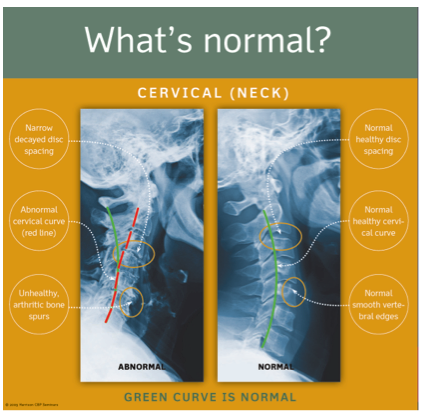
Figure 1. ©Copyright Harrison CBP Seminars. Reprinted with permission.

Literature Review Linking Lordosis to Disorders

            Multiple investigations have been published seeking to understand the association, correlation, or the predictive value of an altered cervical lordosis in different health conditions compared to normal controls. To this end, the majority of these studies have found correlation and predictive validity of the lateral cervical radiographic alignment to a variety of health related conditions including:

  1. Acute and chronic neck pain.2-5
  2. Headaches.5-8
  3. Mental health status.9
  4. Whiplash associated disorders (WAD).10-18
  5. Degenerative joint disease (DJD).19-30
  6. Temporal mandibular joint disorders.31
  7. Range of motion and segmental motion patterns.32-34
  8. Respiration syndromes.35-39
  9. Radiculopathy.40,41
  10. Increased probability for soft tissue injury under impact and inertial loads.42-46

Oppositely, a few investigations have found that the lateral cervical alignment measurements do not correlate to and predict the findings in the above 10 categories.47-52 However, many of these investigations have been found to be internally flawed and detailed reviews of these studies have been performed.53-57 Thus, it should be obvious that the number (45 studies listed above) and the quality of investigations finding a correlation between the lateral cervical radiographic alignment and the conditions in the above 10 categories is superior to the few negative correlation studies. Over the past 20 years, I personally have concluded that the lateral cervical radiographic alignment has positive correlation and predictive validity for the above 10 categories of spine disorders and health conditions.

In Office Methods vs. At Home Care

Historically, the Chiropractic profession has a long history of interest in attempting to improve or correct alterations in cervical lordosis. Problematically, though many outcome investigations have been performed using a variety of chiropractic procedures, most of the traditional methods of Chiropractic procedures have been shown to have limited success at restoration of the cervical lordosis. Still, taken as a collective whole, these reports indicate that patients benefit by reduced pain, improved range of motion, decreased disability levels, and increased health status following chiropractic procedures that improve the cervical lordosis to near normal values.58-85

  • §  In Office Cervical Extension Traction Methods

According to the literature, Chiropractic BioPhysics® or CBP® Technique cervical extension traction procedures are the best available methods for conservatively, consistently and statistically, improving the cervical lordosis. This 'best' evidence exists as 4 clinical control trials (3 non-randomized and 1 randomized) where cervical extension traction treatment methods were added to and compared against various other chiropractic and physical procedures in treated patients versus control group populations.58-61 From this data, extension traction procedures have been found to produce average lordosis corrections between 7° (in more severely injured population) and 18° (in typical chronic neck pain populations) following approximately 36 treatment sessions over the course of 9-12 weeks duration.

Though cervical extension traction procedures can be considered part of the standard of care for rehabilitation of the cervical lordosis; its wide spread implementation into Chiropractic practices has yet to occur. The likely reasons for this lack of widespread implementation for extension traction is multi-factorial and would include: increased square footage office space needed, increased staff to support implementation, increased patient time in the office, and lack of wide spread technical training for proper applications with indications and contraindications for the various methods. See Figure 2. for in office traction methods.

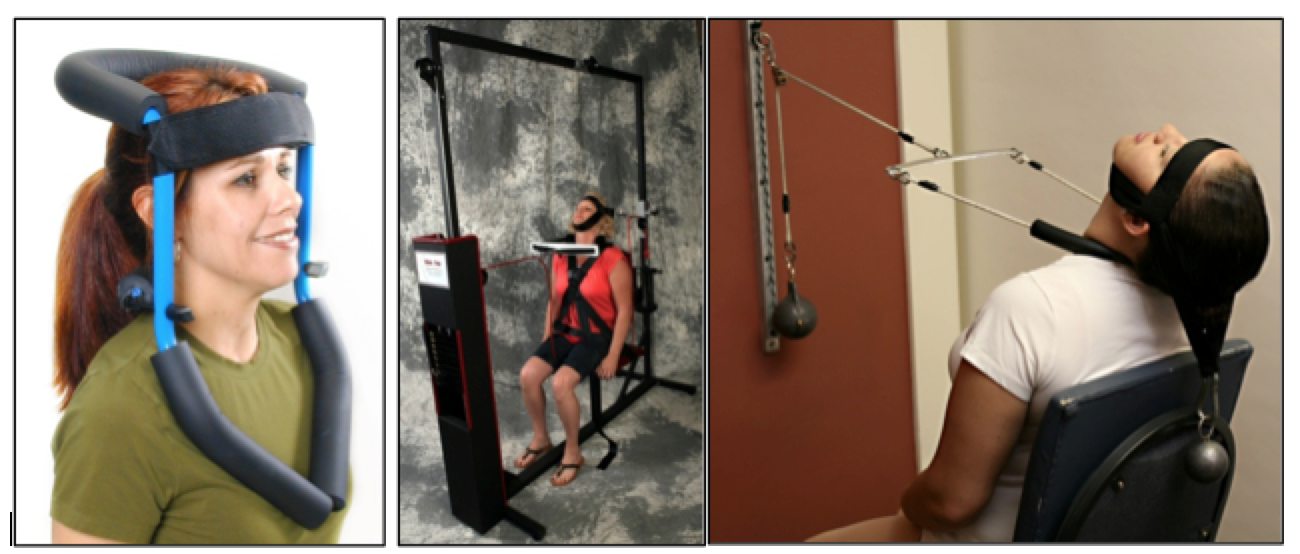
Figure 2. Various in office cervical extension traction methods. ©Copyright Harrison CBP Seminars. Reprinted with permission.

  • §  Home Corrective Orthotics

The use of home corrective orthotic (cervical curve traction) devices as a supplementation to in office treatment programs aimed at rehabilitation of abnormal cervical curvatures has a considerable history in Chiropractic practice. The use of 'at home' cervical extension traction orthotics would seemingly solve several of the key issues with implementation of in office traction methods. Home devices tend to be easier for the patient to use, they are less cumbersome, they are more affordable, and they are likely to be more tolerable. However, at least three main concerns with 'at home' based cervical orthotics must be acknowledged:

There are several different types of home based cervical lordosis corrective orthotics. In Figure 3. a couple of the more popular devices are depicted. Below, I've elected to focus on the cervical denneroll orthotic as it is one of the most applicable, easy to use, and effective (when used properly) home based orthotics today.

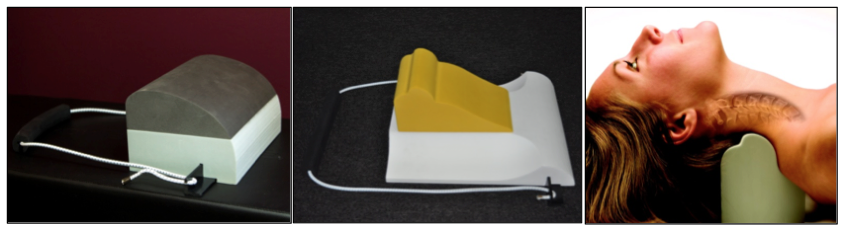
Figure 3. Various at home cervical extension traction orthotics: posture regainer, compression extension unit, and cervical denneroll. ©Copyright Harrison CBP Seminars. Reprinted with permission.

The Denneroll Cervical Orthotic

            The cervical Denneroll orthotic device is a simple, yet complex, pillow-like device engineered with curves, angles, and ridges extrapolated, in part, from the CBP evidence based cervical spinal model. Adrian Dennewald, DC (Denneroll Industries in Sydney, Australia) is the developer and owner of the Denneroll orthotic line. In 2008, Dr. Adrian partnered with Chiropractic BioPhysics in an effort to expand the Denneroll product line, to develop proper indications and contraindications for patient care, and to research-test the effectiveness of the Denneroll in improving the cervical lordosis and patient conditions.

            Personally, I was interested in the Cervical Denneroll device as a solution for a low-stress, comfortable mirror-image® traction orthotic to supplement CBP in office care at home.

            To date, the cervical Denneroll, has been tested in a number of case reports and 2 randomized clinical trials. It's been found to improve the cervical lordosis in different patient populations by 7°-14°.86-91 Thus, the Denneroll orthtoic has been shown to be able to effectively improve the abnormal lordosis of the cervical spine in properly selected cases. Today, the cervical Denneroll products are used worldwide by over 5000 Chiropractors from North America and Australia to the UK, Europe, Asia, and several other international locations.

  • Indications for the Denneroll

The Denneroll currently comes in 3 sizes (adult large, adult medium, and pediatric or small) and can be used in many patient conditions and cervical curve configureations. There are three primary placements of the Denneroll cervical orthotic device shown in Figures 4-6. The Denneroll placement should be consistent with both the shape of the cervical curve and the amount/type of sagittal head translation correction that is desired.

  • Upper thoracic/lower cervical placement- C7-T2 

This placement of the Denneroll will cause significant posterior head translation, it will increase the upper thoracic curve, and increase the overall cervical lordosis. Specifically, this placement should be used for straightened or kyphotic lower cervical segments with loss of upper thoracic kyphosis and anterior head translation of ≤ 40mm. See Figure 4.

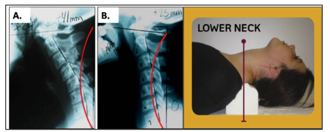
 Figure 4. Abnormal cervical curvatures that fit the inclusion criteria for the application of the Denneroll corrective orthotic in the lower cervical region. These spines must have: • Normal or a mild loss of the upper thoracic kyphosis; • Loss of the lower cervical curve (with or without kyphosis); • Anterior head translation of approximately ≤ 40mm. ©Copyright Harrison CBP Seminars. Reprinted with permission.

Mid-low cervical placement - C4-C6.  This placement of the Denneroll will cause slight posterior head translation; however if the larger Denneroll device is used on a small statured individual then it will create some anterior head translation. The cervical spine should have straightened or kyphotic mid cervical regions (apex of the curve). See Figure 5. In cases with significant posterior head translation, as in Figure 5A, the large Denneroll orthotic should be used and a towel can be placed under the Denneroll to increase the height if needed.

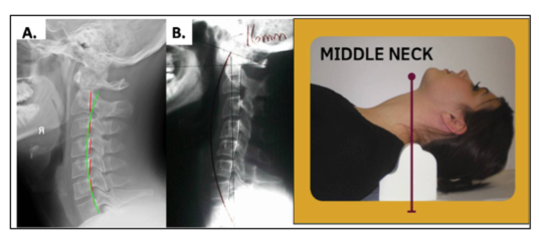
Figure 5. Abnormal cervical curvatures that fit the inclusion criteria for the application of the Denneroll corrective orthotic in the middle cervical region. These spines must have: • Normal or a loss of the upper thoracic kyphosis; • Straightening or apex at the mid-cervical curve; • Slight anterior head translation of approximately ≤ 15mm; • In B with Posterior head translation the LARGE Denneroll should be used with a small towel under it to increase height. ©Copyright Harrison CBP Seminars. Reprinted with permission.

Upper to mid cervical placement- C2-C4. This placement of the Denneroll is used for posterior head translation with straightened or kyphotic mid-upper cervical curves. This position allows extension bending of the upper cervical segments while causing slight anterior head translation. See Figure 6. In cases like Figure 6A with significant posterior head translation, where the posterior vertebral bodies are behind the ideal red curved line,7 the large Denneroll orthotic should be used. While in Figure 6B, the small Denneroll should be used.

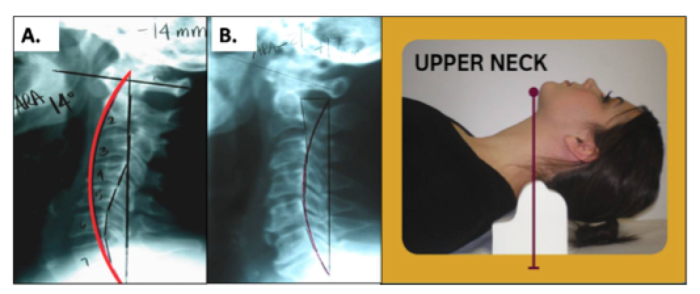
Figure 6. Abnormal cervical curvatures that fit the inclusion criteria for the application of the Denneroll corrective orthotic in the upper cervical region. These spines must have: • Close to normal lower cervical curvature; • Straightening or apex at the C2-C4-cervical segments; • In B, normal head translation of approximately ≤ 15mm. Here the SMALL Denneroll is used to not create anterior head posture; • In A with Posterior head translation the LARGE Denneroll should be used to create anterior head translation. ©Copyright Harrison CBP Seminars. Reprinted with permission.

  • Contra-indications for the Denneroll:

            Quite simply put, no spine orthotic is indicated or should be used in every case presentation. There are both known and proposed risks for extension traction and extension positioning procedures in spine care. The treating -prescribing clinician should perform an examine in every case and perform proper tolerance testing with the patient prior to releasing the patient to use the Denneroll device at home.

            Here's a basic proposed list of contra-indications for Denneroll orthotic patient use:

  1. Moderate to severe mid to upper thoracic hyper-kyphosis;
  2. Large, rigid anterior head translations that does not reduce with extension;

            For a more complete list of contraindications for the Denneroll device, please consider the cervical Denneroll training DVD series available at this link:  http://chiropractic-biophysics.myshopify.com/collections/denneroll-traction-devices/products/denneroll-box-set-all-5-dvds-cervical-thoracic-lumbar-compression-extension-and-scoliroll-training-videos

Simple Case Report for Understanding Home Care Implementation.

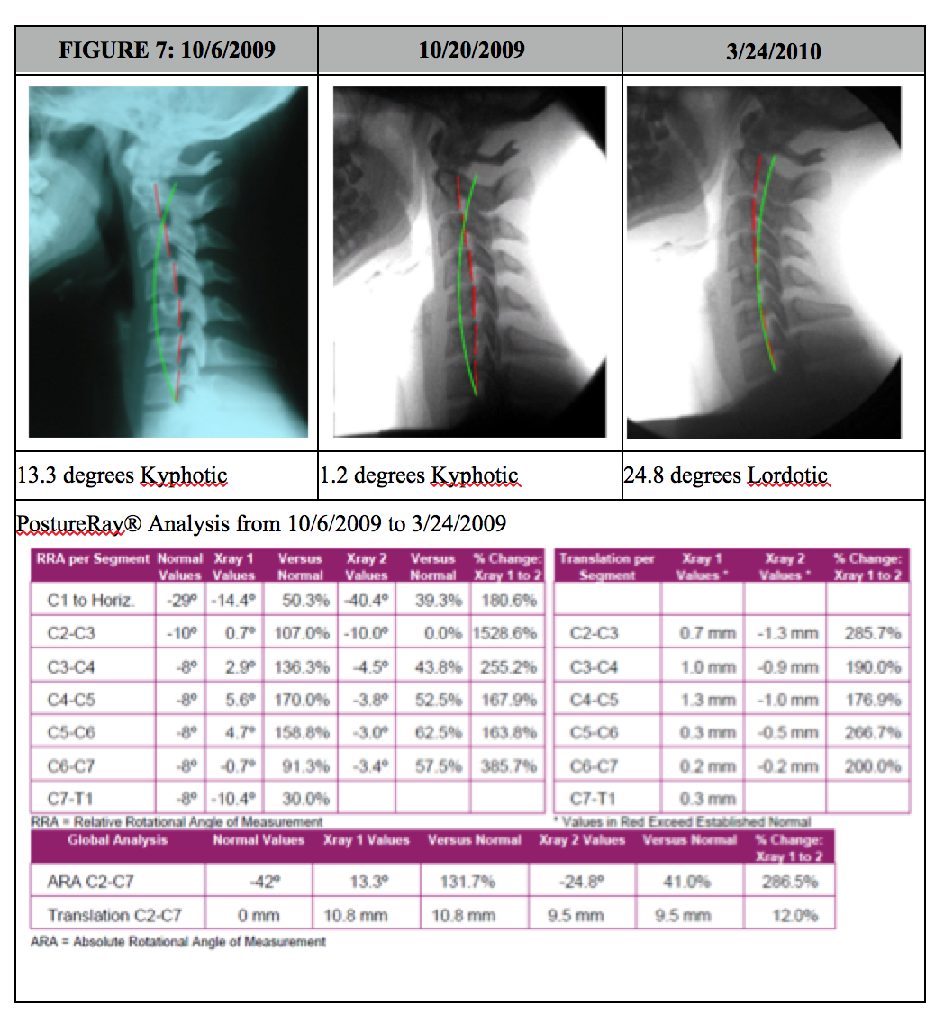
            In this simple / brief case presentation, we have a female that was involved in a frontal collision crash. The subjective complaints were typical complaints seen with whiplash injuries, such as neck pain, sclerotome pain referral to lower neck and upper thoracic spine from probable facet joint injury, headaches, etc. In this case, the patient elected simply not to perform in office traction due to time constraints. In office care consisted of initial coarse of acute care diversified adjusting for 6 visits, then CBP Mirror Image® drop table and instrument based adjustments and Exercises. The patient having such a magnitude of kyphosis, was started as soon as possible with the Denneroll orthotic (on her 7th visit) starting 2x/day at 1 minute and building up to two sessions of 10 minutes, once in morning, and again once at night. Once this goal was achieved, after 2 weeks (4 weeks after injury), she was placed on 1 session of use per day working up to 20 minutes daily. Her initial x-ray was performed on 10/6/2009 then the next post was actually only 2 weeks later, this time on a Digital Motion X-Ray (DMX), dated 10/20/2009.  DMX was chosen as she persisted with headaches and any evidence of ligamentous laxity can be documented. Notice in just 2 weeks of use, the cervical kyphosis is starting to reduce! After 40 sessions of home use, and 36 visits total treatments, with her symptoms and outcome studies showing her nearing pre-injury status, she was prescribed another follow up DMX. The changes on this final follow up x-ray (3-24-2010) were quite amazing as evidenced below in Figure 7.


A Recommendation for In Office & At Home Exercise Warm Up

            It is usually more difficult to re-establish a cervical lordosis in patients that present with a kyphotic cervical curvature and moderate to advanced degenerative joint disease (DJD). These patients usually complain of chronic cervical pain, muscle rigidity and restricted motion. Many of these patients spend much of their day in cervical flexion or anterior head translation and have lost the capacity to truly extend and move their cervical spines. Long term relief for these patients is generally not possible without some form of effective structural and soft tissue rehabilitation. 

            It is for the above reasons that we typically will recommend that patient performs a series of strengthening and flexibility exercises for their cervical spine prior to performing either in office cervical extension traction or at home Denneroll cervical extension traction. Most often we use the Pro-Lordoic Neck Exerciser™ developed by Dr. Don Meyer of California. This device was modified after the cervical neck strap used and taught for this exercise by myself in the CBP Cervical Rehab Seminars for the past several years. Typically we will have the patient perform various forms of cervical exercises using this exercise band for approximately 5-10 minutes prior to performing or using cervical extension traction devices. The Pro-Lordotic exerciser is shown in Figure 8. 

A simple series of exercises with this band are shown on the below youtube links; however, it should be obvious that the treating clinician should select the proper exercises for the individual patient:

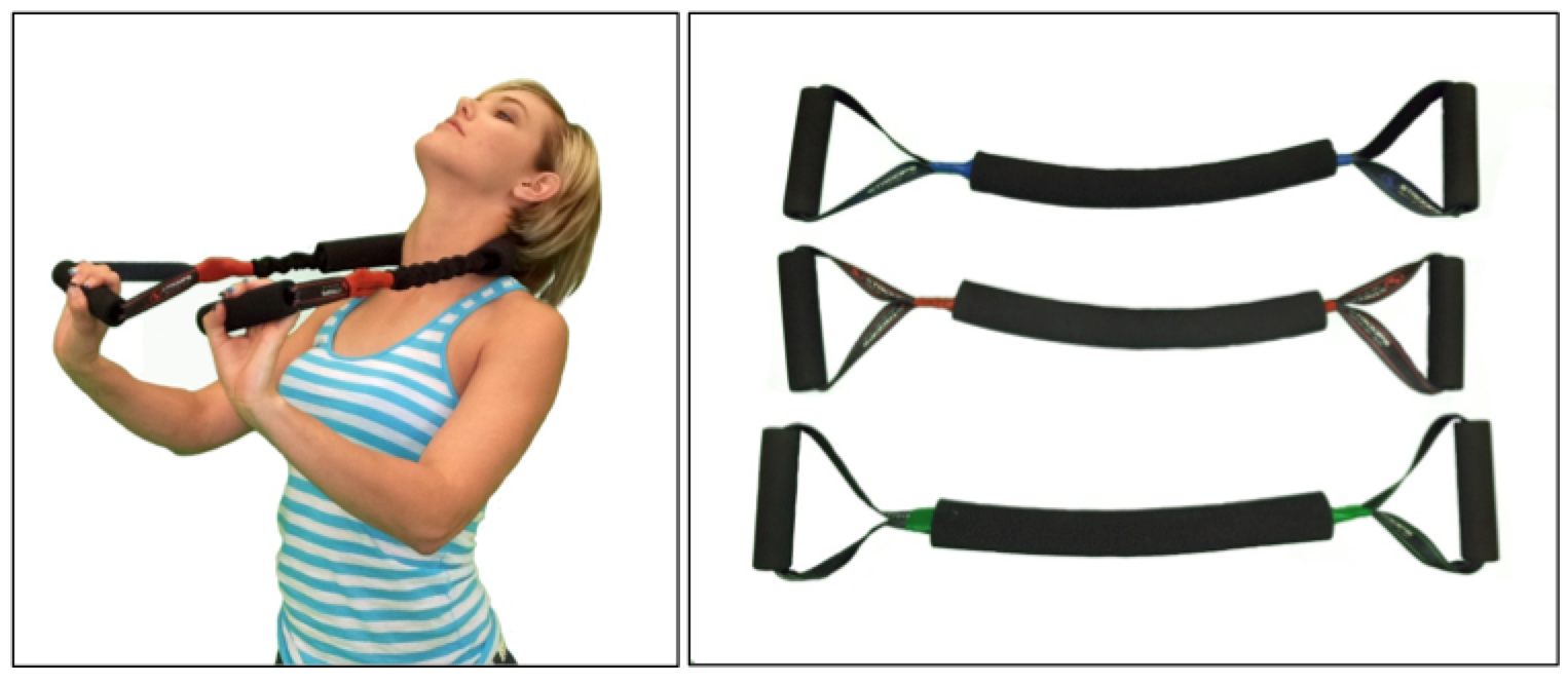
Figure 8. The Pro-Lordotic Neck Exerciser™ is a progressive resistance neck exercise device that tractions the normal lordosis into the cervical spine while active extension exercises of the entire cervical spine are performed during the five minute, structural/postural corrective, home or in-office treatment session. For product ordering information see the following link: http://chiropractic-biophysics.myshopify.com/collections/exercise-equipment/products/pro-lordotic-neck-exerciser ©Copyright Harrison CBP Seminars. Reprinted with permission.

SUMMARY

            Discussions of the cervical lordosis has a long history in the spine literature. While nothing is without controversy, the majority of past and present research reports indicate that the cervical lordosis plays a pivotal role in human health, many spine disorders, and several health disorders. While in office treatment programs combining cervical extension traction procedures should be considered the gold standard for consistent, predictable improvements in patients suffering from abnormalities of the cervical lordosis, at home based corrective cervical spine orthotics should be implemented as well. The cervical Denneroll is one of the most applicable, easy to use, most cost effective, and outcome effective home based cervical extension orthotics on the market today. Clinicians should be aware of the indications and contraindications for at home usage of this device. I hope this presentation assists in your delivery of effective patient intervention in the office and with supplementation of at home devices.


Note:

For more information on the Denneroll Orthotic, please visit http://www.idealspine.com/cervical-dennerol/

For information on becoming a denneroll provider in the USA / Canada please visit http://chiropractic-biophysics.myshopify.com/pages/signup.


References

1.         Harrison DD, et al. Spine 1996; 21: 667-675.

2.         Harrison DD, et al. Spine 2004; 29:2485-2492.

3.         McAviney J, Schulz D, Richard Bock R, Harrison DE, Holland B. Determining a clinical normal value for cervical lordosis. J Manipulative Physiol Ther 2005;28:187-193.

4.         Jochumsen OH. The curve of the cervical spine. The ACA Journal of Chiropractic 1970; August IV:S49-S55.

5.         Choudhary Bakhtiar S; Sapur Suneetha; Deb P S. Forward Head Posture is the Cause of 'Straight Spine Syndrome' in Many Professionals. Indian J Occupat and Environmental Med 2000 (Jul); 4 (3): 122—124.

6.         Nagasawa A, Sakakibara T, Takahashi A. Roentgenographic findings of the cervical spine in tension-type headache. Headache 1993;33:90-95.

7.         Braaf MM, Rosner S. Trauma of the cervical spine as a cause of chronic headache.  J Trauma 1975;15:441-446.

8.         Vernon H, Steiman I, Hagino C. Cervicogenic dysfunction in muscle contraction headache and migraine: A descriptive study. J Manipulative Physiol Ther 1992;15:418-29.

9.         Mears DB. Mental disease and cervical spine distortions. The ACA Journal of Chiropractic 1965; September, pages:13-16,44-46.

10.       Kai Y, Oyama M, Kurose S, et al. Traumatic thoracic outlet syndrome. Orthop Traumatol 1998;47:1169-1171.

11.       Giuliano V, Giuliano C, Pinto F, Scaglione M. The use of flexion and extension MR in the evaluation of cervical spine trauma: initial experience in 100 trauma patients compared with 100 normal subjects. Emergency Radiology 2002;9:249-253.

12.       Giuliano V, Giuliano C, Pinto F, Scaglione M. Soft tissue injury protocol (STIP) using motion MRI for cervical spine trauma assessment. Emergency Radiology 2004;10:241-245.

13.       Marshall DL, Tuchin PJ. Correlation of cervical lordosis measurement with incidence of motor vehicle accidents. ACO 1996;5(3):79-85.

14.       Norris SH, Watt I. The prognosis of neck injuries resulting from rear-end vehicle collisions. J Bone and Joint Surgery 1983;65-B:608-611.

15.       Hohl M. Soft-tissue injuries of the neck in automobile accidents. J Bone and Joint Surgery 1974;56-A:1675-1682.

16.       Zatzkin HR, Kveton FW. Evaluation of the cervical spine in whiplash injuries. Radiology 1960;75:577-583.

17.       Kristjansson E, et al. Is the Sagittal configuration of the cervical spine changed in women with chronic whiplash syndrome? A comparative computer-assisted radiographic assessment. JMPT 2002;25:550-555.

18.       Griffiths HJ, Olson PN, Everson LI, Winemiller M. Hyperextension strain or "whiplash" injuries to the cervical spine. Skeletal Radiology 1995; 24(4):263-6.

19.       Yoon T, Natarajan R, An H, et al. Adjacent disc biomechanics after anterior cervical diskectomy and fusion in kyphosis. Presented at Cervical Spine Research Society, Charleston, SC, Nov. 30-Dec. 2, 2000.

20.       Harrison DE, Harrison DD, Janik TJ, Jones EW, Cailliet R, Normand M. Comparison of axial flexural stresses in lordosis and three buckled configurations of the cervical spine. Clin Biomech 2001;16:276-284.

21.       Harrison DE, Jones EW, Janik TJ, Harrison DD. Evaluation of axial and flexural stresses in the vertebral body cortex and trabecular bone in lordosis and two sagittal cervical translation configurations with an elliptical shell model. J Manipulative Physiol Ther 2002;26:391-401.

22.       Matsunaga S, Sakou T, Sunahara N, et al. Biomechanical analysis of buckling alignment of the cervical spine: predictive value for subaxial subluxation after occipitocervical fusion. Spine 1997; 22: 765-71.

23.       Matsunaga S, Sakou T, Taketomi E, Nakanisi K. Effects of strain distribution in the intervertebral discs on the progression of ossification of the posterior longitudinal ligaments. Spine 1996;21:184-189.

24.       Katsuura A, et al. Kyphotic malalignment after anterior cervical fusion is one of the factors promoting the degenerative process in adjacent intervertebral levels. Eur Spine J 2001;10:320-324.

25.       Matsunaga S, et al. Significance of occipitoaxial angle in subaxial lesion after occipitocervical fusion. Spine 2001;26:161-165.

26.       Matsunaga S, Sakou T, Nakanisi K. Analysis of the cervical spine alignment following laminoplasty and laminectomy. Spinal Cord 1999;37:20-24.

27.       Vavruch L, Hedlund R, Javid D, Leszniewski W, Shalabi A. A prospective randomized comparison between the Cloward Procedures and a carbon fibre cage in the cervical spine: a clinical and radiological study. Spine 2002; 27:1694-1701.

28.       Borden AGB, Rechtman AM, Gershon-Cohen J. The normal cervical lordosis. Radiology 1960;74:806-810.

29.       Harrison DD, Harrison DLJ. Pathological stress formations on the anterior vertebral body in the cervicals. In: Suh CH, ed. The proceedings of the 14th annual biomechanics conference on the spine. Mechanical Engineering Dept., Univ. of Colorado, 1983:31-50.

30.       Yu JK. The relationship between experimental changes in the stress-strain distribution and the tissues structural abnormalities of the cervical column Zhonghua Wai Ke Za Zhi. 1993 Aug;31(8):456-9.

31.       D'Attilio M, Epifania E, Ciuffolo F, Salini V, Filippi MR, Dolci M, Festa F, Tecco S. Cervical lordosis angle measured on lateral cephalograms; findings in skeletal class II female subjects with and without TMD: a cross sectional study. Cranio. 2004 Jan;22(1):27-44.

32.       Panjabi MM, Oda T, Crisco JJ, Dvorak J, Grob D. Posture affects motion coupling patterns of the upper cervical spine. J Orthop Res 1993;11:525-536.

33.       Takeshima T, Omokawa S, Takaoka T, Araki M, Ueda Y, Takakura Y. Sagittal alignment of cervical flexion and extension: Lateral radiographic analysis. Spine 2002;27:E348-355.

34.       Miller JS, Polissar NL, Haas M. A radiographic comparison of neutral cervical posture with cervical flexion and extension ranges of motion. J Manipulative Physiol Ther. 1996 Jun;19(5):296-301.

35.       Ozbek MM, Miyamoto K, Lowe AA, Fleetham JA. Natural head posture, upper airway morphology and obstructive sleep apnoea severity in adults. Eur J Orthod 1998;20:133-143.

36.       Tangugsorn V, Skatvedt O, Krogstad O, Lyberg T. Obstructive sleep apnoea: a cephalometric study, part I. Cervico-craniofacial skeletal morphology. Eur J Orthod 1995;17:45-56.

37.       Hellsing E. Changes in the pharyngeal airway in relation to extension of the head. European J Orthodontics 1989;11:359-365.

38.       Kuhn D. A Descriptive Report of Change in Cervical Curve in a Sleep Apnea Patient: The Importance of Monitoring Possible Predisposing Factors in the Application of Chiropractic Care. JVSR 1998 Vol 3, No. 1, p 1-9.

39.       Dobson, GJ.; Blanks, RHI.; Boone, WR.; Mccoy, HG.; Cervical Angles in Sleep Apnea Patients: A Retrospective Study. JVSR 1999; 3(1): 9-23.

40.       Ferch RD, Shad A, Cadoux-Hudson TA, Teddy PJ. Anterior correction of cervical kyphotic deformity: effects on myelopathy, neck pain, and sagittal alignment. J Neurosurg Spine. 2004;100(1):13-19.

41.       Harwant S. Relevance of Cobb method in progressing sagittal plane spinal deformity. Med J Malaysia. 2001 Dec;56 Suppl D:48-53.

42.       Stemper BD, Yohanandan N, Pintar FA. Effects of abnormal posture on capsular ligament elongations in a computational model subjected to whiplash loading. J Biomechanics 2005;38:1313-1323.

43.       Frechede B, Saillant G, LaVaste F, Skalli W. Risk of injury of the human neck during impact: role of geometrical and mechanical parameters. Paper A29; Presented at the European Cervical Spine Research Society Annual Meeting; 2004 Porto, Portugal, May 30-June 5.

44.       Oktenoglu T, Ozer AF, Ferrara LA, Andalkar N, Sarioglu AC, Benzel EC. Effects of cervical spine posture on axial load bearing ability: a biomechanical study. J Neurosurg (Spine 1) 2001; 943:108-114.

45.       Swartz EE, Floyd RT, Cendoma M. Cervical spine functional anatomy and the biomechanics of injury due to compressive loading. J Athletic Training 2005;40(3):155-161.

46.       Maiman DJ, Yoganandan N, Pintar FA. Preinjury cervical alignment affecting spinal trauma. J Neurosurg. 2002 Jul;97(1 Suppl):57-62.

47.       Gay RE. The curve of the cervical spine: variations and significance. J Manipulative Physiol Ther 1993;16:591-594.

48.       Mamairas C, Barnes MR, Allen MJ. “Whiplash injuries” of the neck: a retrospective study. Injury 1998;19:393-396.

49.       Haas M, Taylor JAM, Gillete RG. The routine use of radiographic spinal displacement analysis: A dissent. J Manipulative Physiol Ther 1999;22:254-59.

50.       Gore DR, Sepic SB, Gardner GM. Roentgenographic findings of the cervical spine in asymptomatic people. Spine 1986;11:521-524.

51.       Peterson CK, et al. Prevalence of hyperplastic articular pillars in the cervical spine and relationship with cervical lordosis. J Manipulative and Physiol Ther 1999;22:390-394.

52.       Li YK, Zhang YK, Zhong SZ. Diagnostic value on signs of subluxation of cervical vertebrae with radiological examination. J Manipulative Physiol Ther 1998; 21(9):617-20.

53.       Harrison DE, Harrison DD, Troyanovich SJ. Reliability of Spinal Displacement Analysis on Plane X-rays: A Review of Commonly Accepted Facts and Fallacies with Implications for Chiropractic Education and Technique. J Manipulative Physiol Ther 1998;21:252-66.

54.       Harrison DE, Harrison DD, Troyanovich SJ. A Normal Spinal Position, Its Time to Accept the Evidence. J Manipulative Physiol Ther 2000;23: 623-644.

55.       Harrison DE. Counter-point article-A Selective Literature Review, Misrepresentation of Studies, & Side Stepping Spine Biomechanics Lead to an Inappropriate Characterization of CBP Technique. AJCC January 2005.

56.       Harrison DE. Counter-point article-A Selective Literature Review, Misrepresentation of Studies, & Side Stepping Spine Biomechanics Lead to an Inappropriate Characterization of CBP Technique. Part II. AJCC April 2005.

57.       Harrison DE, Haas JW, Harrison DD, Janik TJ, Holland B. Do Sagittal Plane Anatomical Variations (Angulation) of the Cervical Facets and C2 Odontoid Affect the Geometrical Configuration of the Cervical Lordosis? Results from Digitizing Lateral Cervical Radiographs in 252 neck pain subjects. Clin Anat 2005; 18:104-111.

58.       Harrison DD, Jackson BL, Troyanovich SJ, Robertson GA, DeGeorge D, Barker WF. The Efficacy of Cervical Extension-Compression Traction Combined with Diversified Manipulation and Drop Table Adjustments in the Rehabilitation of Cervical Lordosis. J Manipulative Physiol Ther 1994;17(7):454-464

59.       Harrison DE, Cailliet R, Harrison DD, Janik TJ, Holland B. New 3-Point Bending Traction Method of Restoring Cervical Lordosis Combined with Cervical Manipulation: Non-randomized Clinical Control Trial. Arch Phys Med Rehab 2002; 83(4): 447-453.

60.       Harrison DE, Harrison DD, Betz J, Janik TJ, Holland B, Colloca C. Increasing the Cervical Lordosis with CBP Seated Combined Extension-Compression and Transverse Load Cervical Traction with Cervical Manipulation: Non-randomized Clinical Control Trial. J Manipulative Physiol Ther 2003; 26(3): 139-151.

61.       Ibrahim M, Aliaa AD, Ahmed A, Harrison DE. THE EFFICACY OF CERVICAL LORDOSIS REHABILITATION FOR NERVE ROOT FUNCTION, PAIN, AND SEGMENTAL MOTION IN CERVICAL SPONDYLOTIC RADICULOPATHY Physiotherapy 2011; 97:supplement 1: 846-847.

62.       Khorshid  KA, Sweat RW, Zemba DA, Zemba BN. Clinical Efficacy of Upper Cervical Versus Full Spine Chiropractic Care on Children with Autism: A Randomized Clinical Trial. JVSR March 9, 2006, pp 1-7.

63.       Wallace HL, Jahner S, Buckle K, Desai N. The relationship of changes in cervical curvature to visual analog scale, neck disability index scores and pressure algometry in patients with neck pain. Chiropractic: J Chiropractic Res Clin Invest 1994; 9:19-23.

64.       Troyanovich SJ, Harrison DD, Harrison DE. A Review of the Validity, Reliability, and Clinical Effectiveness of Chiropractic Methods Employed to Restore or Rehabilitate Cervical Lordosis. Chiropr Tech 1998; 10(1): 1-7.

65.       Alcantara J, Heschong R, Plaugher G, Alcantara. Chiropractic management of a patient with subluxations, low back pain and epileptic seizures. J Manipulative and Physiol Ther 1998;21:410-418.

66.       Alcantara J, Plaugher G, Thornton RE, Salem C. Chiropractic care of a patient with vertebral subluxations and unsuccessful surgery of the cervical spine. J Manipulative and Physiol Ther 2001;24:477-482.

67.       Alcantara J, Steiner DM, Gregory Plaugher and Joey Alcantara .Chiropractic management of a patient with myasthenia gravis and vertebral subluxations.  J Manipulative Physiol Ther 1999;22:333–40.

68.       Alcantara J, Plaugher G, Van Wyngarden DL. Chiropractic care of a patient with vertebral subluxation and Bell's palsy. J Manipulative Physiol Ther. 2003 May;26(4):253.

69.       Araghi HJ. Juvenile Myasthenia Gravis: A Case Study in Chiropractic Management (1993 Proceedings)  http://www.icapediatrics.com/reference-journals.php#.

70.       Araghi HJ Post-traumatic Evaluation and Treatment of The Pediatric Patient with Head Injury: A Case Report (1992 Proceedings) http://www.icapediatrics.com/reference-journals.php#.

71.       Kessinger RC, Boneva DV. Case study: Acceleration/deceleration injury with angular kyphosis. J Manipulative and Physiol Ther 2000; 23:279-287.

72.       Bastecki A, Harrison DE, Haas JW. ADHD: A CBP case study.  J Manipulative Physiol Ther 2004; 27(8): 525e1-525e5.

73.       Ferrantelli JR, Harrison DE, Harrison DD, Steward D. Conservative management of previously unresponsive whiplash associated disorders with CBP methods: a case report.  J Manipulative Physiol Ther 2005; 28(3): e1-e8.

74.       Haas JW, Harrison DE, Harrison DD, Bymers B. Reduction of symptoms in a patient with syringomyelia, cluster headaches, and cervical kyphosis: A CBP® case report. J Manipulative Physiol Ther 2005; 28(6):452.

75.       Colloca CJ, Polkinghorn BS. Chiropractic management of Ehlers-Danlos Syndrome: A report of two cases. JMPT 2003;26:448-459.

76.       Coleman RR, Hagen JO, Troyanovich SJ, Plaugher G. Lateral cervical curve changes receiving chiropractic care following a motor vehicle collision: A retrospective case series. J Manipulative Physiol Therap 2003;26:352-355.

77.       Morningstar, MW.  Cervical hyperlordosis, forward head posture, and lumbar kyphosis correction: a novel treatment for mid-thoracic pain.  J Chiropr Med  2003 Sept;(2:3):111-115.

78.       Morningstar, MW.  Cervical curve restoration and forward head posture reduction for the treatment of mechanical thoracic pain using the Pettibon corrective and rehabilitative procedures.  J Chiropr Med  2002 Sept;(1:3):113-115.

79.       Morningstar, M.W.; Strauchman, M.N.; Weeks, D.A.; Spinal Manupulation and Anterior Headweighting for the Correction of Forward Head Posture and Cervical Hypolordosis: A Pilot Study. J Chiropr Med 2003; 2(2):51-54.

80.       Pierce VP. Results I. Dravosburg, PA: CHIRP, Inc., 1981.

81.       Reynolds, C.; Reduction of Hypolordosis of the Cervical Spine and Forward Head Posture with Specific Adjustment and the Use of a Home Therapy Cushion. Chiropractic Research Journal 1998; 5(1):23-7.

82.       Gary Knutson, DC and Moses Jacob, DC. Possible manifestation of temporomandibular joint dysfunction on chiropractic cervical x-ray studies. J Manipulative Physiol Ther: JAN 1999(22:1) Page(s) 32-37.

83.       Moore MK. Upper crossed syndrome and its relationship to cervicogenic headache. J Manipulative Physiol Ther: JUL/AUG 2004(27:6).

84.       Dobson GJ. Structural Changes in the Cervical Spine Following Spinal Adjustments in a Patient with Os Odontoideum: A Case Report. JVSR August 1996, Vol 1, No. 1, p 1-12.

85.       Moore MK. Upper crossed syndrome and its relationship to cervicogenic headache. J Manipulative Physiol Ther: JUL/AUG 2004(27:6).

86.       Paris B, Harrison DE. Restoration of an Abnormal Cervical Lordosis Using the DENNEROLL: A CBP® Case Report. American Journal of Clinical Chiropractic (ISSN 1076- 7320) 2010; April Vol.20 (2):14,26. http://www.chiropractic-biophysics.com/clinical_chiropractic/2010/4/12/restoration-of-an-abnormal-cervical-lordosis-using-the-denne.html

87.       Ferrantelli JR. BioPhysics Insights: The Denneroll Orthotic. American Journal of Clinical Chiropractic (ISSN 1076-7320) 2010; July Vol.20 (3): 13-14. http://www.chiropractic-biophysics.com/clinical_chiropractic/2010/9/12/the-denneroll-orthotic-i-didnt-believe-it-till-i-tried-it.html

88.       Ferrantelli JR, Harrison DE. Denneroll Combined with Pope 2-Way Aids Patient Suffering from Chronic Whiplash Associated Disorders & Advanced S.A.D.D. American Journal of Clinical Chiropractic (ISSN 1076-7320) 2010; Oct. Vol.20 (4):13,14. http://www.chiropractic-biophysics.com/clinical_chiropractic/2010/10/22/denneroll-combined-with-pope-2-way-aids-patient-suffering-fr.html

89.       Boyd C, Harrison DE. CBP Chiropractors: We Must Practice What We Teach American Journal of Clinical Chiropractic (ISSN 1076-7320) 2012: http://www.chiropractic-biophysics.com/clinical_chiropractic/2012/4/1/cbp-chiropractors-we-must-practice-what-we-teach.html

90.       Does improvement towards a normal cervical sagittal configuration aid in the management of lumbosacral radiculopathy: a randomized controlled trial. In Review for possible publication.

91.       The Effect of Normalizing the Cervical Sagittal Configuration for the Management of Cervicogenic Dizziness: A 1-Year Randomized Controlled Study. In Review for possible publication.
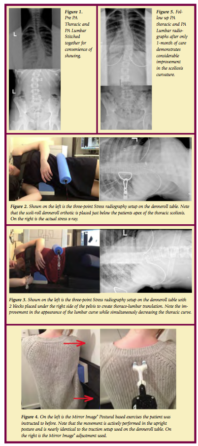
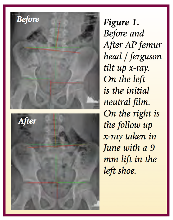
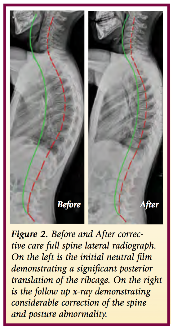
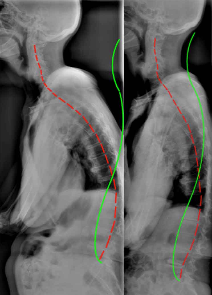
 

Sunday
Jan202013

3-Point Bending Traction for Scoliotic Curvatures Using the New 3-D Denneroll Traction System: A Case Report

INTRODUCTION

In a study from 1893 regarding scoliosis treatment, Bradford and Brackett,1, stated, “there is not only nothing irrational in the method of treatment by forcible mechanical correction when feasible, but it is manifest that when shortened ligaments in spinal curvatures are situated so that they serve as a check to muscular action.”1 They continue, “when they [ligaments] are strong enough to withstand muscular action, gymnastics [exercises] alone are inadequate as a system of correction.” 1 Bradford and Brackett’s1 mechanical traction protocol required patients to undergo traction for a half-hour daily. Because this study was done prior to the invention of x-ray, reported results were not very accurate. More than a century later, CBP® researchers and clinicians have found agreement with Bradford and Brackett that exercises should be combined with short duration, high-force mechanical traction in order to obtain the most effective results in scoliosis reduction.

  • CBP's Mirror Image® Traction for Scoliosis

The traction employed by the CBP® practitioner for scoliosis management requires critical reasoning and a thorough understanding of the displacements of the spine and posture. Generally speaking this traction is of the 3-point-bending type of load application or a transverse load applied at the apex of the curve with and without lateral bending, axial rotation, or other movements depending on the specific case. The traction set-up must always be performed in a pre-determined optimum sequence of movements using stress x-rays to guide the decision making process. Mirror Image® traction sessions and duration should be a minimum of:

  • At least 3-5 times per week. If the patient will traction more than 1 time per day this would be beneficial as long as the patient is not becoming overly painful from the increased frequency of treatment.
  • Traction duration should be 20-30 minutes. The patient starts with 2-3 minutes and over consecutive sessions progresses in time.

CASE REPORT       

            The current patient had a history of thoracic pain and had been under chiropractic care for many years which she indicated gave her temporary relief. Now at 13 yrs old, her pain and frequency have worsened over the last 4 months to a stage where she was experiencing daily headaches and thoracic pain rated as severe on a numerical rating scale (7-8 / 10).

  • Initial Radiography
  1. Primary Right Thoracic curve = 43 degrees (see Figure 1).
  2. Secondary Left Lumbar Curve = 28 degrees (see Figure 1).
  • 1st in traction x-ray using the Denneroll Table and the Scoli-Roll Fulcrum System

            The first in-traction x-ray showed that the thoracic spine was well effected however the lumbar spine was bending and under the stress in the incorrect direction (see Figure 2). This showed us that we needed to raise the lumbar spine off the table to help stretch the lumbar spine correctly.

  • 2nd in traction x-ray

            In response to the first in-traction x-ray, we decided to raise the pelvis to a level of +2 (two blocks under the right hip to address the concerns of the lumbar spine translation. You will see in the 2nd in-traction x-ray that raising the pelvis height did not decrease the effects of the ScoliRoll under the thoracic spine. This is obviously achievable due to the downward pressure of the two straps pulling on the thoraco-lumbar spine and upper thoracic region. The specific effects of using the block system to raise the pelvis is really evident when you look at the stress x-ray in figure 3.

From these in-traction x-rays we can accurately assess that the block under the pelvis is best for the patient’s spine. It also shows how x-rays are essential in establishing the best possible traction position.

  • CHIROPRACTIC INTERVENTIONS

            Due to the positive findings of the stress radiographs, the patient was recommended to undergo corrective chiropractic care including Mirror Image traction on the denneroll table, Mirror image adjusting, and Mirror Image Exercises. She was seen for 3 x week for 1-month (with a couple of interruptions) and was advised on doing home exercises on the days she was not being treated in the office.

  •  Mirror Image® Exercises and Adjustments

            We believe that both postural based exercises and adjustments are vital in consolidating the benefits of the effective spine stretching using the denneroll 3-point bending traction table. During the patient's exercise, neurological stimulation was added by impulsing the spine during her exercise movements; thus turning the exercise into the adjustment.

After 5 weeks and 13 sessions, we can see the corrective improvements in the patient’s spine. The patient’s symptoms have been reduced 90%. Thus, she is symptomatically doing very well and began improving after her 1st session and has reported no symptoms at all for the last 3 weeks.

  • 5 weeks-Follow up Radiography

            A one month follow up radiographs of the thoracic and lumbar spines were obtained to identify if the recommended and applied treatment was having the desired effect. Obviously scoliosis of this magnitude might require more frequent and increased numbers of sessions. However, only a follow up radiograph can truly determine what extent more care or different care is required.

            A remarkable reduction of the AP Thoracic scoliotic curve was identified from 41 degrees down to 28 degrees on the post (a 13° net improvement). Similarly, the AP Lumbar curvature demonstrated improvement. See Figure 5.

SUMMARY

            This case presents the initial successful reduction of a primary thoracic scoliosis in an adolescent female with a history of chronic pain. After 5 weeks and 13 sessions, we can see the corrective improvements in the patient’s spine. The patient’s symptoms have been reduced 90%. We believe the results are due to the combined effect of the Mirror Image treatment methods including the 3-point bending traction employed using the 3-D Denneroll Traction Table. The patient is continuing care and perhaps a future article will address her response.

References

  1. Bradford EH, Brackett EG. Treatment of lateral curvature by mean of pressure correction. 1893.

Sunday
Jan202013

Kinesiology Tape for Postural Control

INTRODUCTION

            As chiropractors, sometimes our efforts become frustrating if our careful attention to adjusting, stretching and strengthening, is undermined by patients resuming the same postural flaws that landed them in our offices in the first place. How exciting would it be to have a sticky, stretchy little assistant that reminded our patients for 2-5 days about positional awareness? Enter elastic therapeutic tape!

            By now, the vast majority of practitioners have had some exposure to elastic therapeutic tape or “kinesio-tape”, the commonly used brand name of developer Kenzo Kase. Tape companies claim it “reduces muscle soreness, improves function, decreases bruising, and decreases pain” and to some extent, these claims appear to be accurate.

            Anything that touches our body’s biggest organ, the skin, has a cutaneous mechanoreceptor effect that stimulates receptors to enhance body kinesthesia or movement awareness. By stimulating large skin mechanoreceptors, kinesiology tape can also downgrade painful stimuli from the nociceptors, which decreases pain perception.

            Early and persistent reasoning suggested that using the tape in an “origin to insertion”, or “muscle action” methodology, best serves to support/stimulate external body areas. While this approach probably makes the most intuitive sense to medical practitioners as it follows anatomical “rules of engagement”, emergent theories, which consider entire postural muscle groups, are making a strong case.

            Dr. Steven Capobianco, chiropractor and developer of the Fascial Movement Taping (FMT) method argues, kinesiology taping should be “based on the obvious yet largely overlooked concept of muscles acting as a chain… the body’s integration of movement via multi-muscle contractions as a means of connecting the brain to the body’s uninterrupted fascial web in order to enhance rehab and athletic performance via cutaneous (skin) stimulation. By taping movement rather than muscles, FMT has demonstrated greater improvement in both patient care and sport performance.

            Dr. Capobianco is not alone in this line of thinking. Leading fascia researcher, Robert Schleip PhD, underscores movement and its role in pain and dysfunction1. Additional support for this model comes from Thomas Myers in his ground-breaking book, “Anatomy Trains”2. He offers a template to assess, treat, and manage body-wide motor dysfunction based on myofascial meridians, and movement impairment.  

            Physical Therapy professor, Heather Murray, makes a strong case for the use of elastic therapeutic tape in those who maintain abnormal postures of the head and neck (i.e. in the work place). Her team conducted a pilot study3, which seemed to suggest that taping for scapular protraction could maintain better posture and decrease perception of pain.

            Recent research indicates that kinesiology tape has a greater stimulatory effect with compromised tissue (due to injury or fatigue due to poor posture). Thedon, et al4 conducted a study to evaluate body sway in individuals with and without tape. They found that the tape showed very little change in the uncompromised condition, but when the subjects were fatigued, the tape provided an added stimulatory effect to the skin helping to compensate for the loss of information fed to the brain from the muscles and joints. For the pain and performance community, this study provides insight into an “auxiliary” system, such as the skin, to augment treatment and training outcomes.

            A 2012 study5 of 32 surgeons, showed a statistically significant reduction in neck and low back pain (using Oswestry Low Back Disability Index and Neck Disability Index) and functional performance (using neck and low back range of motion scores) with the use of kinesiology tape during surgery. This may have far-reaching implications for other jobs/activities where sustained positions result in musculoskeletal pain.

            Let’s look at a common complaint where chiropractors can utilize elastic therapeutic tape to enhance postural control (awareness). Kinesiology tape is commonly used in chiropractic offices to enhance scapular retraction, alleviating upper extremity discomfort caused by internally rotating shoulders (especially when the scapula is in a protracted position). The tape, according to Dr. Capobianco’s model, is applied in a functional manner to augment its therapeutic effect. Begin by addressing the neuro-myo-skeletal dysfunction associated with the protracted shoulder girdle (manipulation, myofascial release, movement re-patterning, etc.). Once the patient is able to appreciate an appropriate retracted/depressed scapula, apply the kinesiology tape (see inset) in a manner to, appropriately, stimulate the local receptors once the intended position is lost.

  • Step 1: Manually mobilize/manipulate the thoracic spine and shoulder girdle and associated myofascial chains
  • Step 2: Place patient/athlete into appropriate postural position that centrates the scapula-thoracic segment.
  • Step 3: Tape the local area (see X pattern and H pattern) with “NO” stretch to stimulate local receptors
  • Step 4: Corrective exercises that will help with postural re-education.


SUMMARY  

            Clearly the use of kinesiology tape is popular (millions of users) and the applications are broad (from athletic injuries to edema). Specific evidence for efficacy is scant but growing, and plausible. There are currently no reported dangers associated with using this elastic cotton mesh bandage, and the only significant contraindication is on open wounds. Kinesiology tape breathes well and flexes like a second skin, unlike most braces that act more like abrasive exoskeletons. It withstands sweat and/or water and is by most comparisons a cost-effective treatment modality. While science is unlikely to discover that kinesiology tape is the panacea for all aches, injuries and postural distortions, medical practitioners should keep this tool in the chest due to its vast possibilities in treating patient complaints and as a tool for postural re-education.

References:

1. Schleip R, Muller D. Training principles for fascial connective tissues: Scientific foundation and suggested practival applications. J Body Move Ther 2012;1-13.

2. Myers, T.W. 2009. Anatomy Trains: Myofascial Meridans for Manual and Movement Therapists. New York: Churchill-Livingston.

  1. Effects of Kinesio Taping on Posture and Presence of Upper Extremity Pain

4. Thedon T, et al. Degraded postural performance after muscle fatigue compensated by skin stimulation. Gait Posture, 2011 Apr;33(4) 686–9.


Sunday
Jan202013

Combining Specific Chiropractic Adjusting Techniques with CBP Corrective Care Techniques: Case #2.

INTRODUCTION

            In our previous article (AJCC October 2012)1, we suggested that the astute Chiropractor should become skilled at administering both a segmental specific adjustment technique (such as the Gonstead system) and a full spine corrective technique like Chiropractic BioPhysics (CBP). In this manner, patients will experience the benefits of segmental motion restriction improvement and the restoration of proper full body and spine alignment. The case presented herein attempts to provide further rationale for the combination of these two uniquely distinct, but complimentary full spine Chiropractic Technique systems.

CBP / Gonstead Case Study #2:

            In 2012, an 8 year old male, who had no overt symptom complaints was brought to the author's (T.P) Chiropractic clinic for postural evaluation and a spinal checkup. His parents had noticed significant posture abnormality and were concerned about underlying spinal disorders.

  • Postural Analysis: Posture analysis revealed a significant left thoracic translation, a right lateral flexion of the ribcage, a right posterior pelvic rotation, a suspected left leg length inequality, a large posterior thoracic translation, and a slight forward head translation. 
  • Radiographic Analysis:  
  1. In April of 2012, a modified AP femur head-ferguson view was obtained. On the left of Figure 1, this x-ray is shown. The PostureRay x-ray software system was used to identify and quantify the extent of leg length inequality. A 9mm left leg length inequality was identified with consequent spine abnormality.

2. In April of 2012, a full spine lateral radiograph was obtained. While in June of 2012, a follow up full spine lateral was obtained to document the response to intervention and potential modifications that might be warranted. Figure 2 depicts these full spine radiographs

  • Interventions: A total of 24 treatment sessions over the course of 2 months was utilized in this case.

            For segmental adjusting technique, the treating Chiropractor (TP) exclusively adhered to and utilized the Gonstead Technique system for identified spine subluxations including:

  1. Abnormal temperature differential patterns (nervo-scope / tempo-scope).
  2. Static palpation data indicating the presence of edematous tissue around the injured segment.
  3. A decrease in motion of the segment in question, as compared to the surrounding area.
  4. Palpable muscle spasm or splinting around the area in question.
  5. Visualization of the area (looking for presence of pitting edema, asymmetry in the tissues, etc).
  6. Then, consulting the 3-shot, digitally stitched, AP full spine x-ray and the lateral (2 shot) full spine x-ray to analyze the “foundation principle” and relate this to the examination findings.

            For the full spine and posture corrective care, CBP® mirror image® adjusting, exercise, traction procedures were utilized. Additionally, body weighting using the thoracic belt from Circular Traction was applied 5 times per week for 15 min walking intervals. These procedures were administered on each visit to the patient. Lastly, a full foot lift of 9 mm was administered to the patient and was placed in the left shoe.

  • Mirror Image Adjustments

The patient was administered mirror image adjustments to correct posterior thoracic translation with hyper-kyphosis and anterior head translation with head flexion postures. See Figure 3.

  • Mirror Image Exercises

The patient was administered mirror image® exercises to correct posterior thoracic translation with hyper-kyphosis and anterior head translation postures. See Figure 4.

  • Mirror Image® Body Weighting and Denneroll Traction

      Mirror Image traction care was administered to the patient. The patient received the denneroll traction system to correct the abnormal thoracic translation posture as shown in Figure 5. In addition the patient was instructed in the use of the denneroll thoracic orthotic and was advised to do this 5 times per week at home.

  • Case Outcome

            Subjectively, at the end of 2 months of corrective care, the patient's posture was remarkably improved. NRS = 1-2 / 10. According to the patient's parents, in their own words, "It’s great to see how quickly he has improved. The leveling of his shoulders and hips is apparent and how he carries himself; he is dramatically more upright and balanced".

            Objectively, the patient's follow up full spine radiographs in Figure 2 show remarkable reduction of the spinal displacements.


CONCLUSION

            The authors' opinion is that the patient's improvements were directly related to both the segmental adjusting and spinal corrective procedures applied using CBP Technique. Accordingly, for optimum patient response, traditional Chiropractic adjustments would seem to be enhanced by the addition of spinal corrective procedures as in CBP. In the end, it is the positive response of the patient that should dictate this combined approach of classical Chiropractic care, with more contemporary corrective Chiropractic systems.

References

  1. http://www.chiropractic-biophysics.com/clinical_chiropractic/2012/12/18/combining-specific-chiropractic-adjusting-techniques-with-cb.html


Sunday
Jan202013

Certainty and 2013

Randall Hammett, DC

Private Practice of Chiropractic


INTRODUCTION

            The one consistent thing is change, and I tell you for sure 2013 will be a challenge for many chiropractors. With changing health laws both federal and state changing insurance policies to cover shortages you can expect more financial stress and more focus on documented care with less reimbursement. In my own town I’ve already been notified that several government-backed insurance plans are increasing their deductibles and copayments by vast amounts to cover shortages. The good news is, and there is good news! The health industry has typically been inflation and recession proof. Don’t get me wrong, doctor’s incomes have dropped the last five years and yet it’s been estimated that 5 to 7% of doctor’s incomes have increased. The question is what will your practice hold for you in 2013?        

DISCUSSION

            The last three years have seen unusual solutions to practice problems in chiropractic. One example is the low fee Doc in a box corporate chiropractor office that seemingly undercuts every chiropractor’s fee in their path. As a practitioner for over 33 years I’ve seen many financial games played in the profession and in my experience the low fee high-volume chiropractic offices eventually collapse financially, it’s not if it’s only when. My opinion about your fees is simply charge what you believe you are worth and the true value of the services you provide. One of the things I recommend that you do in 2013 is receive chiropractic care from a colleague near you and pay them their full fee for each treatment. Psychologically, paying out of your own pocket for chiropractic care will put you in touch with the reality of what your patients have to pay and in some cases you’ll find that you’re not charging enough for the services you provide. The keynote for the year is to stay flexible in your business planning and in your practice marketing. Keep in mind that patients will always gravitate to the practice where doctors produce outstanding, fast symptomatic relief with good post pain educational information for patients to decide if they wish to continue with the chiropractic lifestyle.

            Be sure in your practice that you offer patients at least three types of care for their health. Pain relief, corrective care and wellness or maintenance care are typically the three types of care that we offer patients.  An important part to remember is that you must honor what patients choose and not step over your bounds. For example the patient wants relief care for a few visits than honor that and when they are out of pain release them and explain to them that they’re always welcome back if and when the pain returns. Corrective care should be based on strict clinical protocols such as those found in CBP® so that patients can clearly understand what they’re paying for and can easily see the postural x-ray changes you’re providing. Wellness or maintenance care should be recommended for everyone but typically in the chiropractic office only 5 to 7% will follow through, if this is true in your practice then you’re doing a good job and continue with your treatment plans. There has been in the last several years a propensity to base your care on what third-party insurance companies will pay for, and there could be no higher injustice to a patient than treating their wallet instead of their health.

There’s an old saying, you can steal someone’s money and they can earn it back, but if you steal someone’s health they can never replace it. 

SUMMARY

            So, when recommending care to patients, tell them the truth, let them decide what’s important at that time to them and not necessarily to you. Lastly, what’s your plan for 2013 to increase your practice a minimum of 25 to 30%? If you’ve not taken a day to sit down and write out what your plan is for the next twelve months I guarantee you that your practice will decrease and your income will fail. Those of you reading this that got into chiropractic because it was a good career move, or good way to earn income I suggest you become quickly reacquainted with the purpose and philosophy of chiropractic because if you’re in it just for the money you will never survive and you will always struggle financially and emotionally. It amazes me to today how many chiropractors practicing have never read any of the green books, have never attended a chiropractic philosophical seminar like DE in Atlanta. The chiropractors who have survived and thrived in the last 100 years have done so by first providing outstanding results, second by educating their patients as to why they need chiropractic for a lifetime, and third understanding the limits of the care that they provide and the extent to which a chiropractic adjustment, performed correctly can change people’s lives.

Till next time,

Sunday
Jan202013

The Why and How of Practice Outreach

Yurij Chewpa, DC, RFCCSS(C),

Co-Head Coach, Warrior Coaching and Warrior Coaching USA (www.WarriorCoaching.org)

 INTRODUCTION

            At the recent Warrior Coaching Leadership Summit in Toronto, the topic of the various presentations was practice outreach. Specifically, the speakers were discussing "the why" - why do we do outreach, and "the how" - how do we reach out into our communities effectively to attract the sick and lost. What strategies work best?

            The strategies being discussed were screenings, corporate talks, and patient dinners. These three strategies have been around for decades and many falsely assume that they no longer work. Nothing could be further from the truth. While internet marketing, a social media presence, and a great website are crucial in today’s wireless world, they do not replace getting outside of your four walls and personally interacting with the people in your community. Warrior Coaching clients receive hundreds of new patients every month using these tried and true strategies. As a matter of interest, if done correctly they are more effective today then they have ever been in the past. It is not unusual to schedule 50 to 100 great patients at a 2-day screening, or schedule 20 to 30 patients from a 20-minute dinner, or corporate, presentation.

DISCUSSION

            One of the advantages of doing the outreach in person is that the doctor has to wrestle with the question of why they are doing it, and face the fear of getting outside of their four walls. Are they doing this talk to get new patients to build their practice and pay their bills, or are they doing it to save the sick people in their community? The better the answer as to why the doctor is doing the talk, the more people will come in for a checkup. If it is all about the doctor, the potential patients will sense this and they will stay away. If the people have an understanding that the doctor is there to serve them, then that doctor has the capacity to attract as many patients as he can possibly serve.

            Once the doctor knows why they are doing the outreach, they need to know how to do the outreach most effectively. The same doctor can give the same talk to the same group and change the content by five percent and get a 30, 60 or 100-fold increase in follow-through. Likewise, we have had Warriors do screenings and schedule 100 new patients in a weekend, and a chiropractor at the same screening one booth over only schedule 10 patients.

"The why" or "the how"?

            After the presentations at Leadership Summit there was a lot of discussion about what is more important, "the why" or "the how"? Why we do outreach, or how we do outreach? Obviously, both are important, but does either one have an advantage when it comes to building a lifetime, wellness, family, principled practice?

            The why v. how question can be stated in other ways as well: inspiration v. perspiration, delivery v. content, passion v. procedure.

            When it comes to practice, doctors have to have a certain amount of both. Everybody's personalities are unique. Some doctors gravitate towards why, others towards how. Both can be successful, it just looks a little different.

            Here are my observations over the past 27 years of practice and 12 years of coaching. The Why practice tends to have more new patients and better initial conversion, but more early drop outs. This is because the doctor tends to be outgoing and passionate and has no problem attracting new patients, but lacks the procedures to start a patient well and doesn’t have the process to guide them on their path of care. Practice tends to grow fast at the beginning, but plateaus because of lack of structure. This is a practice that tends to have a high missed appointment rate and high dropout rates, but can be high volume because the new patients are ever present. The inmates are running the asylum. By adding structure, this practice is ready to explode.

            The How practice tends to have lower new patients and a slightly lower conversion rate, but a much higher retention rate, because the structure and procedures guide the patient through their months and years of care. Practice growth is slower because the doctor tends to be more fearful of outreach, and therefore new patient numbers are lower. Although practice growth is slow, it is more consistent and steady. The inmates are not running the asylum like they are in the why practice, but it's hard to break into the asylum. By adding more new patients, this practice is ready to explode.

SUMMARY  

            As you can see, one is not better than the other, they are just different. The Why doctor is not mindful of structure and procedure. The How doctor is fearful of outreach. Both need to step into fear to reach their full God-given potential. Both doctors will have trouble sustaining practice growth without investing consistently into both areas. The best scenario is to have a good mix of both. With a good amount of how and why, a practice can have steady, sustainable growth.

            Questions for you to answer after reading this should include: Where do your tendencies fall? Where do you need to be courageous, and where do you need to step into fear?

Sunday
Jan202013

Learn to talk TO a person WITH a subluxated posture at your ROF.

Fred DiDomenico, DC

Practice Coach and Mentor

www.elitecoachingllc.com


INTRODUCTION

         Most management groups teach you scripts of how to talk ABOUT a subluxation to a person at your ROF. What does this imply?  You are talking about the facts of a suluxated posture to a person.  It is as though the patient will understand the facts, see their x-rays and commit to care because it makes sense.  Unfortunately it doesn’t always seem to work that way, because there continues to be people walking out your door who, “just don’t get it.”  Have you noticed talking MORE about the same thing doesn’t make you more effective?  Successful business people don’t use the excuse, “they just don’t get it,” so why should we?

         People make decisions and buy based on an emotional progression of thought and feelings.  There are 5 steps a patient or any person who is going to make an empowered action toward a life change MUST progress through in a specific order. These 5 steps incorporated into your patient management system will raise your percentage of people committing to your spinal corrective programs to SOAR HIGHER than ever before.

  1. They MUST SAY they have a FULL spine problem:

            Entrepreneurs solve people’s problems for money.  This means people MUST know their problem and be very clear.  In a spinal corrective program we correct the WHOLE spine.  Every spinal problem involves the WHOLE spine.  The question is, “Do you have the systems that have the HIGHEST percentage of patients understanding their WHOLE spine needs correcting?”  The next question is, “Do they tell YOU they KNOW their WHOLE spine and posture is weak and subluxated?”  The fact is you can’t tell them. They MUST Say it to you so they will understand.  It’s not what YOU say that matters.  It’s what THEY say that changes their behavior.

2. The MUST say, “I don’t want to be like this anymore.

            Out of our GREATEST crisis comes our greatest breakthrough.  Before people are willing to make a change in their life they MUST want to leave the condition they are in.  They must reach the point of ultimate frustration to change and be ready to move.  Many patients that don’t commit NEVER said, “I don’t want to be like this anymore.”  This is a CRUCIAL EMOTIONAL decision people MUST make to be ready to move in another direction. They must have a strong emotional desire to change or they will stay in their misery, whether it is their pain or their disease.

3. Eliminate ALL other options:

            Once a person makes the emotional commitment to move in another direction, they need guidance as to the proper direction to move.  You don’t achieve this by telling them “What to do.” You achieve this by telling them “What NOT to do.”    Research shows that people with chronic back pain who do only exercise have a greater probability of becoming disabled than if they did nothing at all. This eliminates traditional physical therapy, medical care, yoga, Pilates, gyms, and all the other exercise related excuses, including, “Getting a second opinion.”  If you don’t eliminate all other options, you will receive common objections to care and non-commitment at your ROF.  Elite Coaching provides these answers in systems so the HIGHEST percentage of people commit to corrective care by directing their actions.

4. They MUST know what they DO want:

            Once a person is ready to move in a specific direction they MUST have a goal. Without a goal they do NOT know what they want and will not buy and say, “I have to think about it.”  The problem is you didn’t direct them to have a vision in advance.  “Where there is no vision, people perish,” right out of your office.  Over 80% of people DON’T write goals. Without a vision of what they want they will not have the emotional commitment or a  clear destination to move toward.  For this reason we have patients write 20-30 year health/life goals.  We “coach” them to see the life they desire with GREAT emotional attachment and they also see they can’t live that life if they allow their subuxated spine and posture to remain and progress.  NOW your program becomes the vehicle to get what THEY want.  This is one of the many reasons Elite Coaching clients receive greater value for spinal corrective care.  We teach you how to TRULY “coach” your patients.

5. Prove your correction:

            Show them a post rehab, corrected x-ray of their spine. This is physical proof that builds trust, faith and confidence that proves your result without words.  Their check for your program relates to trust.  When you follow ALL these steps and finish with a post rehab x-ray they lose the ability to object.

            These steps are in a very specific order with intention based on how people act emotionally and make decisions. Follow these steps and you will have the opportunity to help and serve MORE patients in 2013 than EVER BEFORE! Attend the Elite Coaching ROF Boot Camps and create the MOST FULFILLING practice of your life! Call Dr. Fred DiDomenico for more information, 253-851-8353.


Sunday
Jan202013

Business And Practice Tips Secrets to Business Success

Eric Huntington, DC

Co-Owner Developer of the Chiropractic Business Academy

drhuntington@chirobizacademy.com


INTRODUCTION

Why is Business Success a Secret?

            There are many aspects to running a business which will determine its degree success.  From delivering good service to honest dealing with your clientele to motivating staff—many of these things are “known” and  applied broadly in our profession. But there are lesser known and applied business principles that really separate the proverbial men from the boys.  It’s these principles that remain a secret.

  • Secret #1

            You must find out what is needed and wanted by the public, and then figure out how you can promote and deliver that item or service. This is best done by survey, formal or informal. How does this apply to chiropractic practice?  Well, kick your ego to the curb and ask yourself this question, “Does the public want chiropractic?”  I’m not suggesting blasphemy here, I’m suggesting we be literal.  To be more specific, “Does an individual in the public want chiropractic?”  No, of course not—an individual, to be interested in purchasing and receiving the services of a chiropractor, would want the BENEFITS of chiropractic. 

            The benefits of chiropractic are numerous and can include a healthy body, better movement, less pain, better posture, etc.  It’s worth listing out what you see as the benefits a patient receives when under chiropractic care.   This list should be used to help formulate your promotion and technical delivery.

            Secret #1 is that when you organize your promotion and delivery, you must do so keeping in mind what is needed and wanted by the public.  As an example, this is why some of the marketing strategies taught by the Chiropractic Business Academy utilize massage.  By survey, massage is a more  needed and wanted service than chiropractic at the moment.

  • Secret #2

            Secret #2 is that you must build a machine to promote and sell whatever is needed and wanted.  By “build a machine” I mean you must hire or create competent personnel.  Lines and flows must be efficiently organized so daily office traffic runs smoothly through the business.  Training manuals are needed for reference by staff and to assist in training new staff.  Written policy ensures that staff actions are coordinated and predictable.  These are just a few building blocks taught by CBA that can assist in building your machine.

            You know when the machine is built and operating, because it will run without you having to do most of the work.  Depending on how you set it up, you may still work in the practice, wearing the hat of your choice, or you may phase yourself out completely.  That’s up to you.

            At the Chiropractic Business Academy (CBA) we have helped thousands of chiropractors build their machine.  Since we teach business skills, including marketing, sales, staff training, finance, leadership, etc, our program works for any practice style.

  • Secret #3

            Once you have attained your ideal practice scene, you must continually monitor it using proper statistics.  Secret #3 is becoming an expert at looking at what you are doing that works, and improving those things—and also determining what you are doing that is not working and making changes to those areas.

            I think it’s worth noting that more than half of the doctors that contact CBA for practice help are struggling to one degree or another.  Good news is that we can help any doctor who is willing to learn and work.  Our strategies are proven in every state and several countries for almost two decades. 

            Conversely, many doctors miss a huge opportunity because they don’t think to contact us when they are doing well.  This is the optimal time to hire CBA because we can help a doctor identify what is causing their success and strengthen it!  In fact, the biggest practice gains from our program are routinely experienced by clients who were already doing well when they joined CBA. 

  • Secret #4

            Your own happiness in practice may be the most important factor.  You get to define success in your life.  You also get to decide how to measure your progress toward your goals.  So this secret is unique to each of us. You may find it helpful to list out what would make you happy in practice.  For me, my list includes things like:

  1.  
    1. Providing high quality service
    2. Staff driven practice which I don’t work in day to day.
    3. High personal income
    4. Freedom to choose when to work
    5. Freedom to travel

            These are just a few examples from my list, but what is important is that you make your list and go for it! If you are certain that you can achieve you goals without the help of a consultant, that’s great.  If not, give my office a call and we can talk about how CBA can help you get there!

            CBA’s program is made to fit your practice and goals, not the other way around.  Call us and let us know how we can help. We are so sure our program will increase your bottom line that we make that guarantee in our client service agreement.

            Call my office today to schedule a free consultation to learn the exact system we have used to help thousands of chiropractors over the last 15 years! 888-989-0855


Sunday
Jan202013

CBP® NonProfit, Inc. Research Update

Check Out Our Research Reference List Online at: http://www.idealspine.com/cbp-research/

Paul A. Oakley, M.Sc., DC

CBP Research & Instructor

Private Practice New Market, Ontario, Canada


            Chiropractic Biophysics Non-profit, Inc. is a 501(c)(3) nonprofit corporation dedicated to the advancement of chiropractic principles through scientific research. Dr. Don Harrison (deceased) and his second wife Dr. Deanne LJ Harrison (deceased) founded CBP research foundation in 1982; it was registered as CBP Non-Profit, Inc. in 1989 by Dr. Sang Harrison (Don’s 3rd and final life’s love). Through this organization Dr. Don and colleagues have published 140 peer-reviewed spine and Chiropractic research publications. Further, CBP Non-Profit, Inc. has funded many scholarships as well as donated chiropractic equipment to many chiropractic colleges; always trying to support chiropractic advancement and education. Dr. Don Harrison was the acting president of CBP Non-Profit, Inc. since 1982. Currently, Dr. Deed Harrison (Don’s son) is the President of CBP Non-Profit, Inc.

            Results of our studies have been published in prestigious research journals and presented at respected conferences around the world. Your (Chiropractic donations) support enables us to continue important research and gives you a voice in the course our studies take. Join today, either as a regular member or member of the President’s Circle. The result will be better chiropractic techniques, stronger chiropractic practices, and healthier chiropractic patients.

CBP® research consists of studies on a variety of CBP® technique related topics including:

  1. Spine modeling studies evaluating ideal and average human alignment variables,
  2. Spine biomechanics studies analyzing loads, stress, and strains,
  3. Posture modeling studies,
  4. Reliability of measurements and evaluation of patients (x-ray, posture),
  5. Validity of the measurements and evaluation of patients,
  6. Randomized trials evaluating technique outcomes
  7. Non-randomized trials evaluating technique outcomes,
  8. Case series studies evaluating technique outcomes,
  9. Case studies evaluating technique outcomes,
  10. Literature reviews and professional commentaries.