


3-Point Bending Traction for Scoliotic Curvatures Using the New 3-D Denneroll Traction System: A Case Report


INTRODUCTION
In a study from 1893 regarding scoliosis treatment, Bradford and Brackett,1, stated, “there is not only nothing irrational in the method of treatment by forcible mechanical correction when feasible, but it is manifest that when shortened ligaments in spinal curvatures are situated so that they serve as a check to muscular action.”1 They continue, “when they [ligaments] are strong enough to withstand muscular action, gymnastics [exercises] alone are inadequate as a system of correction.” 1 Bradford and Brackett’s1 mechanical traction protocol required patients to undergo traction for a half-hour daily. Because this study was done prior to the invention of x-ray, reported results were not very accurate. More than a century later, CBP® researchers and clinicians have found agreement with Bradford and Brackett that exercises should be combined with short duration, high-force mechanical traction in order to obtain the most effective results in scoliosis reduction.
- CBP's Mirror Image® Traction for Scoliosis
The traction employed by the CBP® practitioner for scoliosis management requires critical reasoning and a thorough understanding of the displacements of the spine and posture. Generally speaking this traction is of the 3-point-bending type of load application or a transverse load applied at the apex of the curve with and without lateral bending, axial rotation, or other movements depending on the specific case. The traction set-up must always be performed in a pre-determined optimum sequence of movements using stress x-rays to guide the decision making process. Mirror Image® traction sessions and duration should be a minimum of:
- At least 3-5 times per week. If the patient will traction more than 1 time per day this would be beneficial as long as the patient is not becoming overly painful from the increased frequency of treatment.
- Traction duration should be 20-30 minutes. The patient starts with 2-3 minutes and over consecutive sessions progresses in time.
CASE REPORT
The current patient had a history of thoracic pain and had been under chiropractic care for many years which she indicated gave her temporary relief. Now at 13 yrs old, her pain and frequency have worsened over the last 4 months to a stage where she was experiencing daily headaches and thoracic pain rated as severe on a numerical rating scale (7-8 / 10).
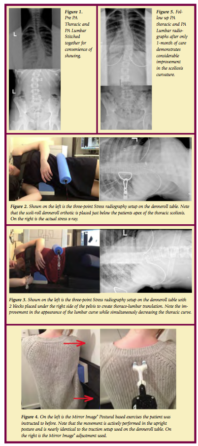
- Initial Radiography
- Primary Right Thoracic curve = 43 degrees (see Figure 1).
- Secondary Left Lumbar Curve = 28 degrees (see Figure 1).
- 1st in traction x-ray using the Denneroll Table and the Scoli-Roll Fulcrum System
The first in-traction x-ray showed that the thoracic spine was well effected however the lumbar spine was bending and under the stress in the incorrect direction (see Figure 2). This showed us that we needed to raise the lumbar spine off the table to help stretch the lumbar spine correctly.
- 2nd in traction x-ray
In response to the first in-traction x-ray, we decided to raise the pelvis to a level of +2 (two blocks under the right hip to address the concerns of the lumbar spine translation. You will see in the 2nd in-traction x-ray that raising the pelvis height did not decrease the effects of the ScoliRoll under the thoracic spine. This is obviously achievable due to the downward pressure of the two straps pulling on the thoraco-lumbar spine and upper thoracic region. The specific effects of using the block system to raise the pelvis is really evident when you look at the stress x-ray in figure 3.
From these in-traction x-rays we can accurately assess that the block under the pelvis is best for the patient’s spine. It also shows how x-rays are essential in establishing the best possible traction position.
- CHIROPRACTIC INTERVENTIONS
Due to the positive findings of the stress radiographs, the patient was recommended to undergo corrective chiropractic care including Mirror Image traction on the denneroll table, Mirror image adjusting, and Mirror Image Exercises. She was seen for 3 x week for 1-month (with a couple of interruptions) and was advised on doing home exercises on the days she was not being treated in the office.
- Mirror Image® Exercises and Adjustments
We believe that both postural based exercises and adjustments are vital in consolidating the benefits of the effective spine stretching using the denneroll 3-point bending traction table. During the patient's exercise, neurological stimulation was added by impulsing the spine during her exercise movements; thus turning the exercise into the adjustment.
After 5 weeks and 13 sessions, we can see the corrective improvements in the patient’s spine. The patient’s symptoms have been reduced 90%. Thus, she is symptomatically doing very well and began improving after her 1st session and has reported no symptoms at all for the last 3 weeks.
- 5 weeks-Follow up Radiography
A one month follow up radiographs of the thoracic and lumbar spines were obtained to identify if the recommended and applied treatment was having the desired effect. Obviously scoliosis of this magnitude might require more frequent and increased numbers of sessions. However, only a follow up radiograph can truly determine what extent more care or different care is required.
A remarkable reduction of the AP Thoracic scoliotic curve was identified from 41 degrees down to 28 degrees on the post (a 13° net improvement). Similarly, the AP Lumbar curvature demonstrated improvement. See Figure 5.
SUMMARY
This case presents the initial successful reduction of a primary thoracic scoliosis in an adolescent female with a history of chronic pain. After 5 weeks and 13 sessions, we can see the corrective improvements in the patient’s spine. The patient’s symptoms have been reduced 90%. We believe the results are due to the combined effect of the Mirror Image treatment methods including the 3-point bending traction employed using the 3-D Denneroll Traction Table. The patient is continuing care and perhaps a future article will address her response.
References
- Bradford EH, Brackett EG. Treatment of lateral curvature by mean of pressure correction. 1893.

 CBP Seminars